Figure 1. Fundus aspects from two patients
homozygous for a MER tyrosine kinase protooncogene (MERTK)
deletion. A-C: 22-year-old female (patient 106); the
central black spot. The central black spot is an artifact to reduce
reflexes from the Zeiss fundus camera. A: In the posterior part
of the retina, near-normal calibrated central arterioles and a
normal-appearing optical nerve head are evident. B: In the
inferotemporal aspect, widespread mottled pigment epithelial atrophy
and heavy pigment aggregates partly sheeting the retinal venules are
seen. C: In the macular region, distinct, nearly circular,
foveal atrophy is present. D: In the left eye from a female age
35 years (patient 232), marked atrophy of the optic papilla,
constricted arterioles, and distinct central pigment atrophy are
present.
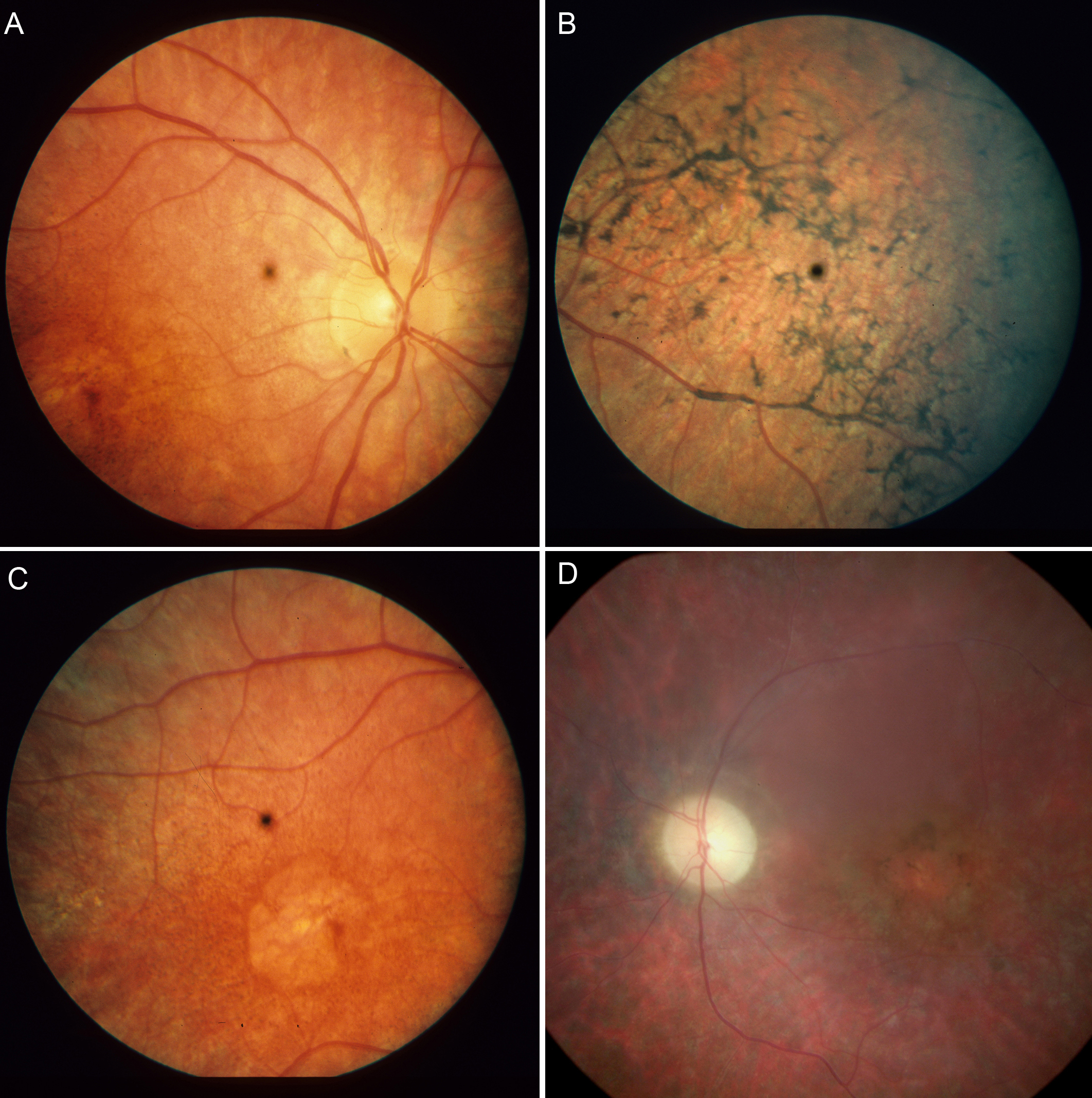
 Figure 1 of Ostergaard, Mol Vis 2011; 17:1485-1492.