Figure 6. Representative western blots showing cytochrome c levels in gup mutants treated with diferuloylmethane. Cytochrome c (cyto c) levels were determined in embryos treated for 6 days. Mitochondrial
and cytosolic fractions were normalized with cox IV or β-actin, respectively. Quantitation of western blot of cytochrome c
localization in larvae is shown directly beneath western blots. Wildtype levels in each fraction are set to 1.0. Wt, wildtype;
gup, untreated mutant larvae, gup+D, mutant plus diferuloylmethane.
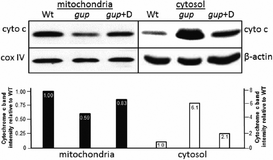
 Figure 6 of
Gregory-Evans, Mol Vis 2011; 17:1473-1484.
Figure 6 of
Gregory-Evans, Mol Vis 2011; 17:1473-1484.