Figure 3. Representative images of drug treatment in gup mutants at 6 dpf. A, C, E, G, I: Enlarged wholemount of the eye, scale=200 µm, and wholemount larvae, scale=500 µm. B, D, F, H, J: corresponding TUNEL stained wholemount of PTU-treated eye and larvae. A, B: Wildtype zebrafish showing normal morphology and minimal apoptosis in the eye and wholemount. C, D: Untreated gup mutants displaying large coloboma in ventral aspect of the eye, TUNEL-positive labeled tissue at the site of the unfused
optic fissure and throughout the whole fish. E, F: gup mutants treated with 5 µM diferuloylmethane (+D) showing small colobomatous defect, minimal TUNEL-positive staining in the
eye, and reduced levels in the whole larvae compared to untreated mutants. G, H: gup mutants treated with 300 µM zVAD-fmk (+V) showing small colobomatous defect, minimal TUNEL-positive staining in the eye,
and reduced levels in the whole larvae compared to untreated mutants. I, J: gup mutants treated with 300 µM zFA-fmk (+F) showing large colobomatous defect and TUNEL-positive staining at the site of the
unfused fissure.
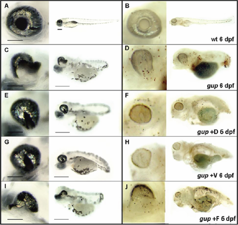
 Figure 3 of
Gregory-Evans, Mol Vis 2011; 17:1473-1484.
Figure 3 of
Gregory-Evans, Mol Vis 2011; 17:1473-1484.