Figure 2. Representative morphology of gup mutants at 48 hpf following drug treatment. Left panel, enlarged image of wholemount eye, scale bar=200 μm. Right panel,
whole embryo morphology, scale bar=500 μm. A: Wildtype control, complete closure of optic fissure. B: Untreated gup mutant, displaying large open optic fissure at ventral aspect of eye. C: gup mutant treated with 5 µM diferuloylmethane, displaying open optic fissure which is smaller in size compared to untreated
mutants. D: gup mutant treated with 300 µM zVAD-fmk, displaying a smaller open optic fissure than untreated mutants. E: gup mutant treated with 300 µM zFA-fmk, displaying a large open optic fissure as in untreated mutants. Optic fissure closure
defect delineated by black dotted line.
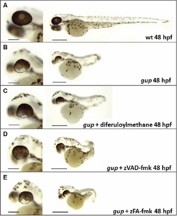
 Figure 2 of
Gregory-Evans, Mol Vis 2011; 17:1473-1484.
Figure 2 of
Gregory-Evans, Mol Vis 2011; 17:1473-1484.