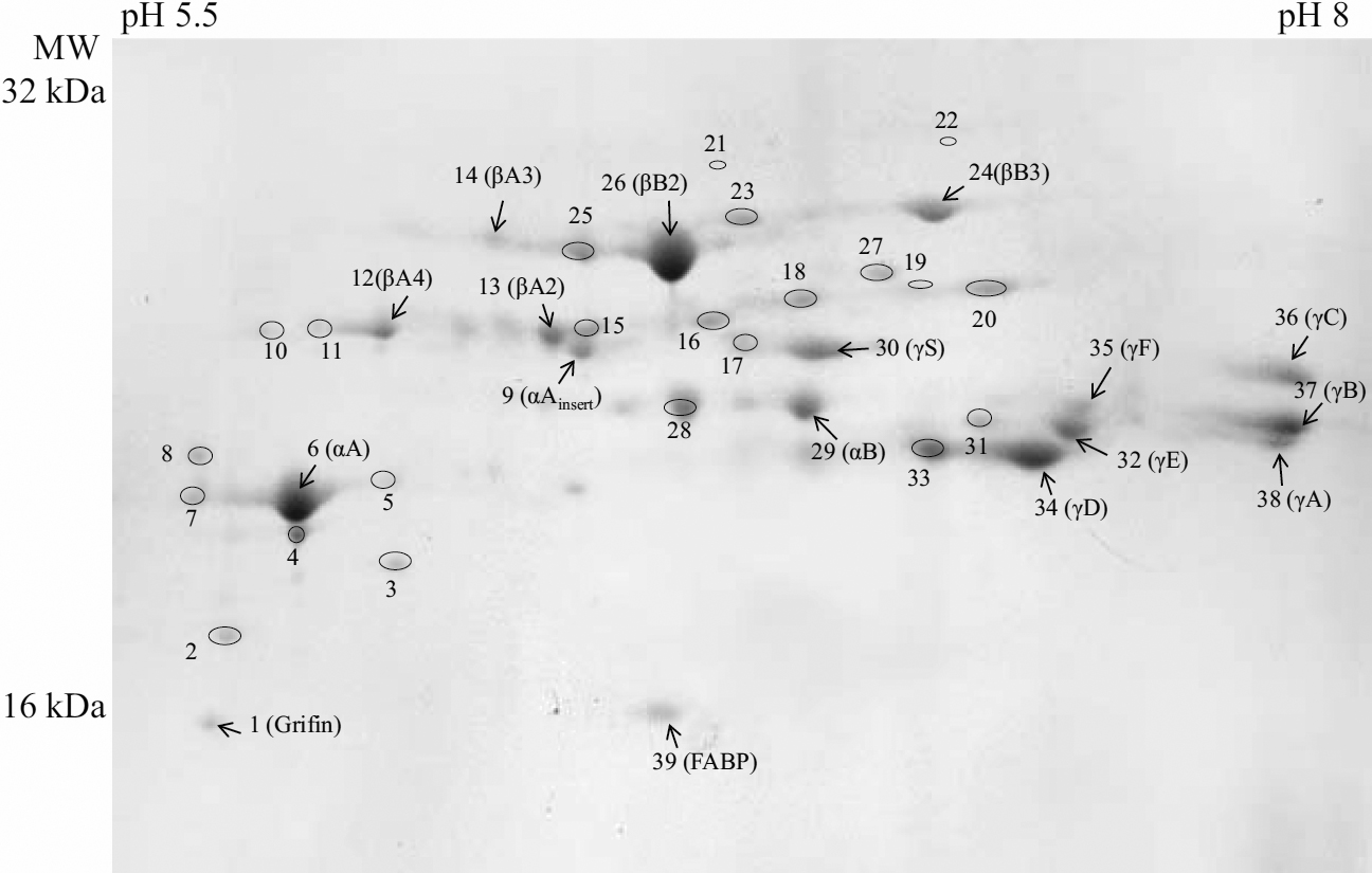
Figure 1. 2-DE gel of the lens proteins of
12-week-old Wistar rat stained with Coomassie brilliant blue R-250.
Arrows indicate the unmodified proteins, and their assignment is shown,
the modified crystallins are given the numbers only: spots 2–5,7,8
correspond to αA-crystallin, spots 10,11 – to βA4-crystallin, spots
15–20 – to βA3-crystallin, spot 22 – to βB1-crystallin, spot 23 – to
βB3-crystallin, spots 26–27 – to βB2-crystallin, spot 28 – to
αB-crystallin, spot 31 – to γE-crystallin and spot 33 – to
γD-crystallin. The assignment of all indicated spots and the sequence
coverage are also presented in
Table 2.
 Figure 1 of Kopylova, Mol Vis 2011; 17:1457-1467.