Figure 3. Electron microscopy of lacrimal
gland. Representative H&E staining and electron microscopy
photographs of non-Sjögren syndrome (case 9; A, B, C)
and
Sjögren syndrome patient (case 4; D, E, F)
were shown. Scale bar indicated 5 μm (B and E) and 2 μm (C
and F). Structure of lacrimal acinar unit was compact and
uniform in non-Sjögren syndrome patients (A and B), but
mild acinar atrophy and fibrosis were observed more frequently in
Sjögren syndrome patients (D and E). High magnification
revealed that structure of mitochondrial cristae (arrows) was severely
damaged and swollen in Sjögren syndrome patient (F) compared to
that in non-Sjögren syndrome patient (C).
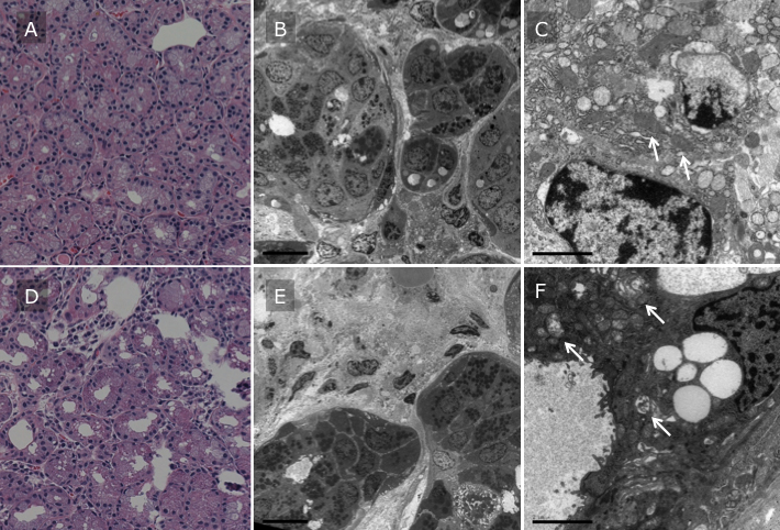
 Figure 3 of Kawashima, Mol Vis 2011; 17:1397-1404.