Figure 2. Immunostaining for progenitor
markers. In non-Sjögren syndrome, p63 (red) was expressed in 2–4 cells
in each acinar unit (A) and all ductal basal cells (B;
case 7). In Sjögren syndrome, p63 was weakly expressed with irregular
pattern. (case 2; F, G). Nucleostemin was expressed
with a similar pattern in non-Sjögren syndrome (case 7; C) and
Sjögren syndrome (case 2; H). Nuclei were counterstained with
DAPI (blue). ABCG2 (red) was expressed in intercellular junction and
cytoplasm in acinar unit (F, I), and weaker in Sjögren
syndrome. Nestin was expressed strongly in some location in Sjögren
syndrome (E, J). Scale bars indicate 50 μm (A-C,
E-H, J) and 20 μm (D, I),
SS=Sjögren syndrome, non-SS=non Sjögren syndrome.
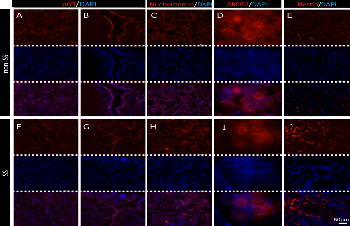
 Figure 2 of Kawashima, Mol Vis 2011; 17:1397-1404.