Figure 4. Exon organization of
xlProminin-1, 2, and 3. Homologous exons of xlProminin-1, 2, and 3 are
aligned. Genes of all three xlProminins are alternatively spliced and
their exon organization is evolutionarily conserved. Constitutive exons
are marked in black. Alternatively spliced exons are marked orange.
Spliced forms identified in cDNA clones are indicated by joining lines.
Translation start sites (ATG) are marked with green lines and the
positions of translational stop codons are marked with white lines.
Note the complex splicing of exons 26b, 27, and 28a, which generates
several distinct isoforms of xlProminin-1. Positions of primers used in
reverse-transcription (RT)-PCR for semiquantitative analysis of mRNA
expression and for alternative splicing are indicated on the diagram.
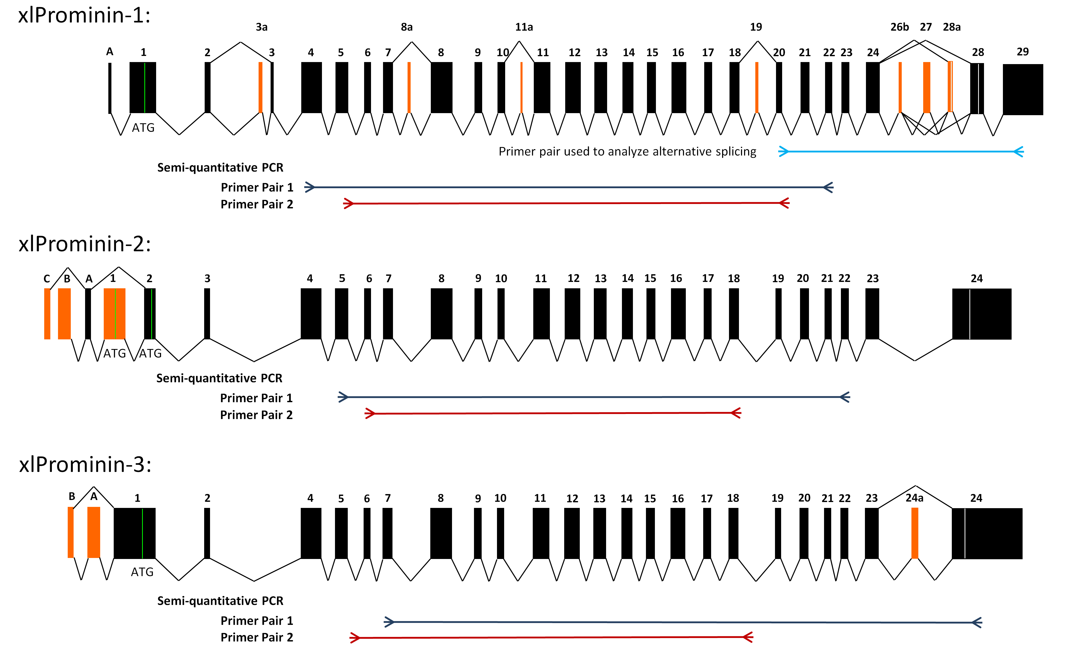
 Figure 4 of Han, Mol Vis 2011; 17:1381-1396.