Figure 2. Morphological changes caused by the addition of anti-human integrin antibodies to human scleral fibroblasts (HSFs) in the
collagen gel lattice. Anti-human integrin antibody (at the concentration 4µg/ml, which was diluted with serum-free DMEM) was
added dropping it onto an HSF-populated collagen gel. Morphological changes of fibroblasts were monitored after 6 h addition
using light microscopy (original magnification 200×). A: mouse anti-human IgG (0.1 mg/ml) was used as a control; B: A mixture of anti-human integrin a1 (4 µg/ml) and anti-human integrin β1 (4 µg/ml) antibodies were added. C: A mixture of anti-human integrin a2 (4 µg/ml), and anti-human integrin β1 (4 µg/ml) antibodies were added.
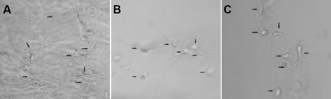
 Figure 2 of
Hu, Mol Vis 2011; 17:1334-1342.
Figure 2 of
Hu, Mol Vis 2011; 17:1334-1342.