Figure 1. Identification of the collagen-binding integrins subtypes expressed in human scleral fibroblasts (HSFs). A: Amplification products representing the integrin α1 (280 bp), integrin α2 (226 bp) and integrin β1 (180 bp) subunits were
detected in HSFs using reverse-transcription PCR. Molecular markers were included for product size comparison. B: The products representing the integrin α1 (130 kDa), integrin α2 (129 kDa), and integrin β1 (88 kDa) subunits were detected
in HSFs using western blot analysis. C-K: Distribution of integrin α1, α2 and β1 in HSFs were observed by indirect immunofluorescence. FITC marked the secondary antibody
(green; 1) and Hoechst33358 dyed the nucleus (blue; 2). The first (1) and second images (2) combined to form the third image
(3). Integrin α1 (C-E), α2 (F-H), and β1 (I-K) were localized in the plasmalemma of HSFs.
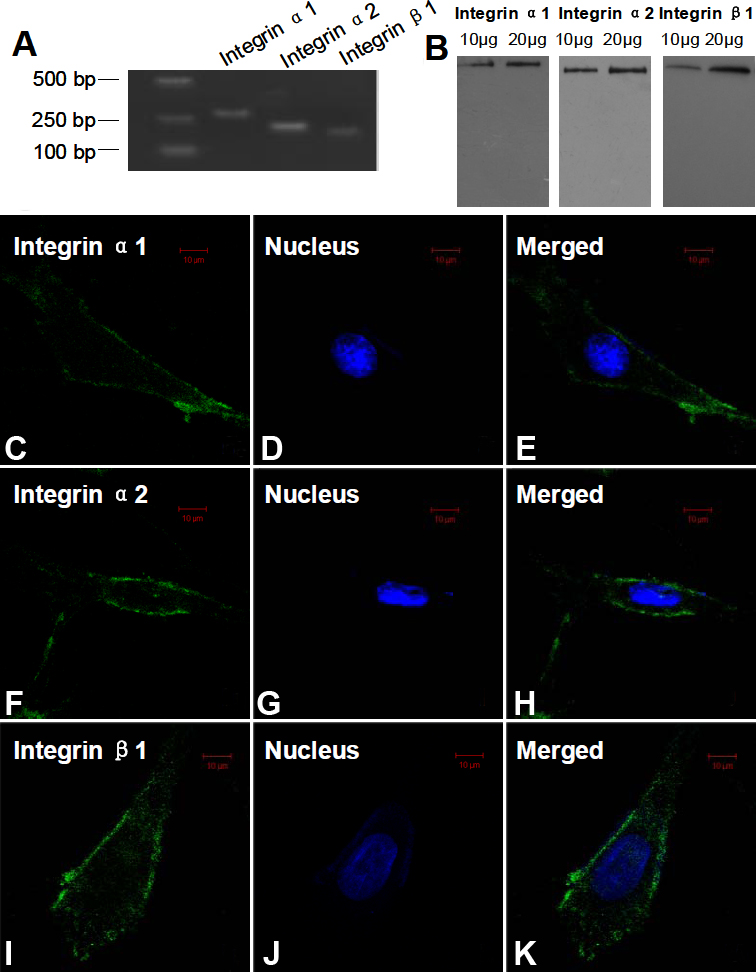
 Figure 1 of
Hu, Mol Vis 2011; 17:1334-1342.
Figure 1 of
Hu, Mol Vis 2011; 17:1334-1342.