Figure 6. SEM images of human corneal
microvilli and mucin network and in standard and EDEV HCE. Scanning
electron microscopy showing HCE epithelium in control HCE condition (A,
B at 20 and 5 μm of magnification) and in EDEV setting at 24 h (C,
D), 48 h (E, F), and 72 h (G, H).
The microvilli in C-H are identified at a different
magnification (2–5 μm). The in vivo impression cytology with SEM on
humans is illustrated in the box on the top of the Figure (microvilli
on the left and mucin network on the right). The reproduction of the
images are authorized by Professor Del Prete (University of Napoli).
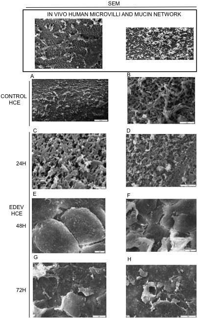
 Figure 6 of Meloni, Mol Vis 2011; 17:113-126.