Figure 5 of
Meloni, Mol Vis 2011; 17:113-126.
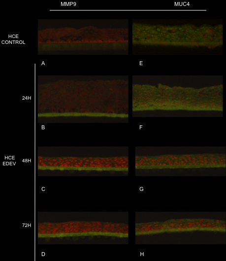
Figure 5.
Immunofluorescence staining of MMP9 (
A
,
B
,
C
,
D
) and MUC4 (
E
,
F
,
G
,
H
) in control and EDEV HCE at 24 h, 48 h, and 72 h. MMP9 and MUC4 are stained in green and red nuclei are stained with propidium iodide.
 Figure 5 of Meloni, Mol Vis 2011; 17:113-126.