Figure 10. Images of microvilli in in
vitro reconstituted corneal epithelium. Scanning electron microscope
images showing Control (A) and EDEV-HCE tissue induced for
24 h (B) with treatments with different tear substitutes (C-Hyalistil®;
D-Etacortilen®; E-Xiloial®; F-TSP
0,5%®; G-Optive®). Two μm magnification.
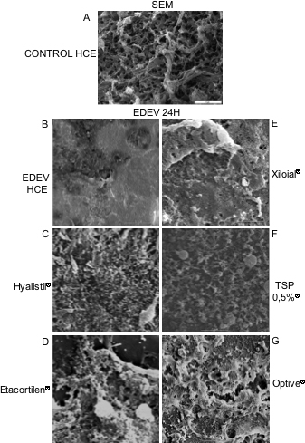
 Figure 10 of Meloni, Mol Vis 2011; 17:113-126.