Figure 1. Histopathological analysis of
the anterior eye segment. A-H: Light micrographs of rat eyes
stained with toluidine blue. A, B: Ciliary body of
control animals. C: Degranulated mast cell (curved arrow) in
the ciliary body and extensive inflammatory cell infiltration in AqH (D),
mainly neutrophils (arrows) at 24 h after endotoxin injection (LPS
24h). E: Intact mast cell (curved arrow) in the ciliary process
and mononuclear phagocytic cells (open arrows) adhered to the iris
surface (F) at 48 h post-injection (LPS 48h). G, H:
Ciliary bodies of animals pretreated with secretagogue c48/80 before
EIU (c48/80 + LPS 24h). Absence of mast cells and very few
transmigrated inflammatory cells (arrow). V: blood vessel. Bars, 10 μm.
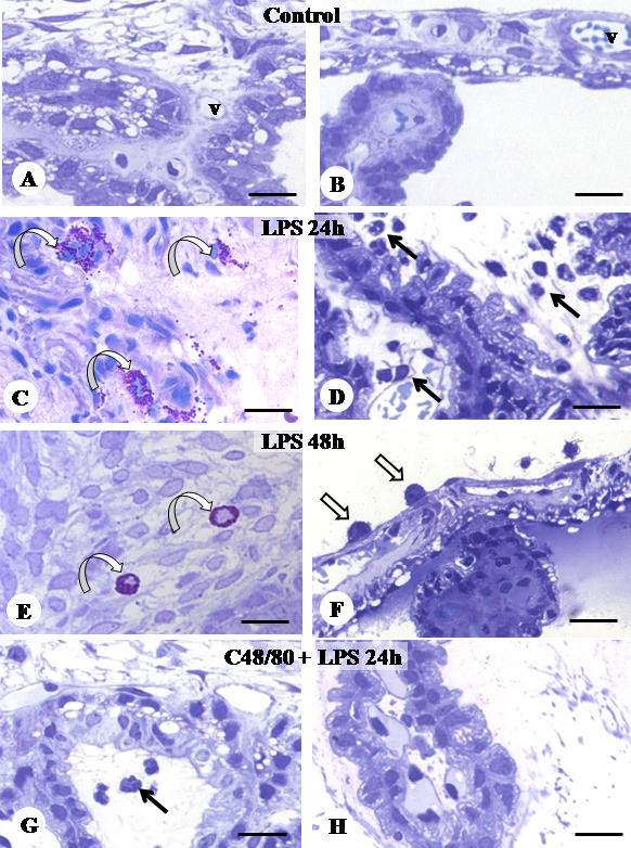
 Figure 1 of Sebastião da Silva, Mol Vis 2011; 17:1310-1319.