Figure 6. Localized expression of Glb1l3
in retinal cell layers. Wild-type retina sections were used to perform
combined in situ hybridization (ISH) and immunohistochemistry (IHC)
using cell type-specific retinal markers of the rods (recoverin), the
cones (cone arrestin; cArr), the horizontal (calbindin) and bipolar
(PKCα) cells of the INL, the Müller cells (GFAP), and the ganglion
cells (Brn3b) of the GCL. As the staining for Brn3b was too weak to be
detected in a combined experiment, two adjacent retina sections were
used for Glb1l3 in situ hybridization and Brn3b
immunohistochemistry. Glb1l3 mRNA was mainly expressed in the
rods, in the cells of the INL—including horizontal and bipolar cells,
and in the ganglion cells. OS, outer segments; ONL, outer nuclear
layer; INL, inner nuclear layer; GCL, ganglion cell layer.
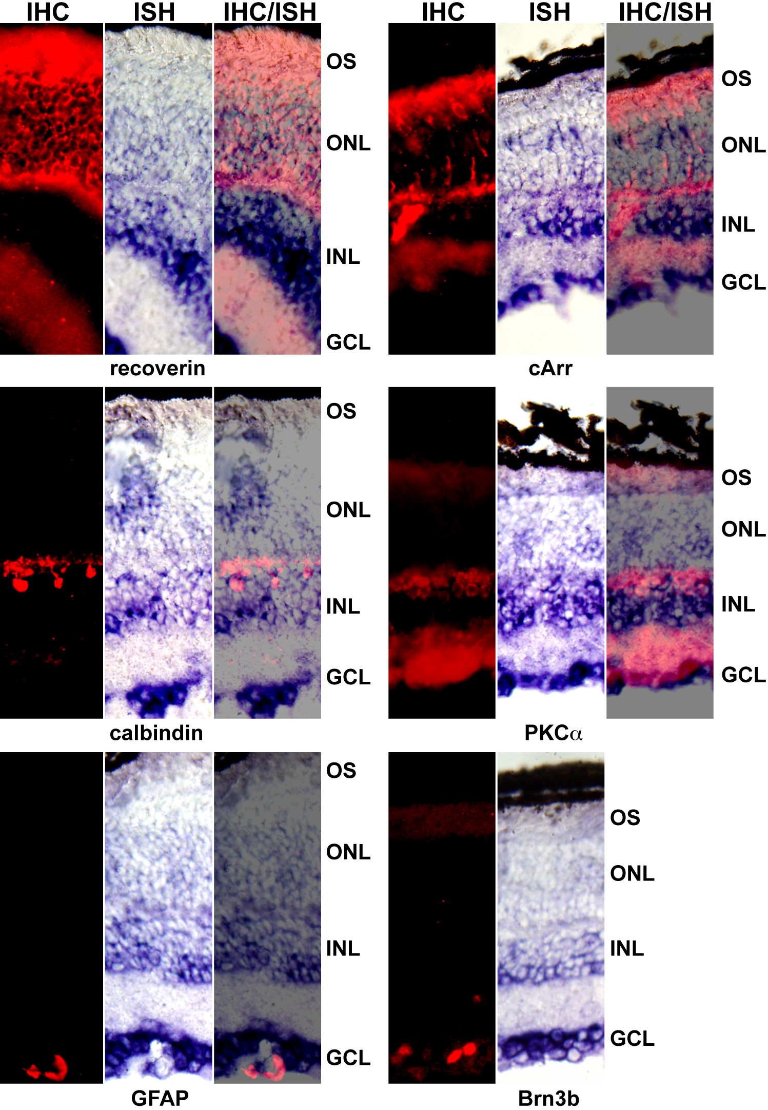
 Figure 6 of Le Carré, Mol Vis 2011; 17:1287-1297.