Figure 2. Hydrogen sulfide
preconditioning–mediated antiapoptotic effects. A: Effect of
hydrogen sulfide (H2S) preconditioning on caspase-3
activation 24 h after unilateral ischemia. Pro-caspase-3 and caspase-3
levels were determined using specific antibodies. Histograms represent
the densitometric ratio of caspase-3 cleavage compared with loading
control (glyceraldehyde 3-phosphate dehydrogenase [GAPDH]). The amount
of pro-caspase-3 and protein loading seemed comparable in all groups
(lanes 1–4). Compared to control, ischemia/reperfusion (I/R) injury led
to a significant cleavage from pro-caspase-3 to active caspase-3 (lane
1 versus 2). H2S preconditioning before I/R injury
significantly reduced cleavage of pro-caspase-3 to caspase 3 (lane 4
versus 2; ***p<0.001). Data are presented as mean±SD of five
experiments. B: Fluorogenic caspase-3 assay (DEVDase assay) of
full retinal protein lysates 24 h after I/R injury. Caspase-3 activity
was low in control eyes (room air) and was not significantly affected
by H2S inhalation in controls. I/R injury increased the
activity (p<0.001 compared with control eye). In contrast,
preconditioning with inhaled H2S significantly reduced
caspase-3 activity in ischemic tissue. Results are given in RFUs. Data
are presented as mean±SD of eight experiments. ***p<0.001 I/R injury
versus H2S + I/R injury.
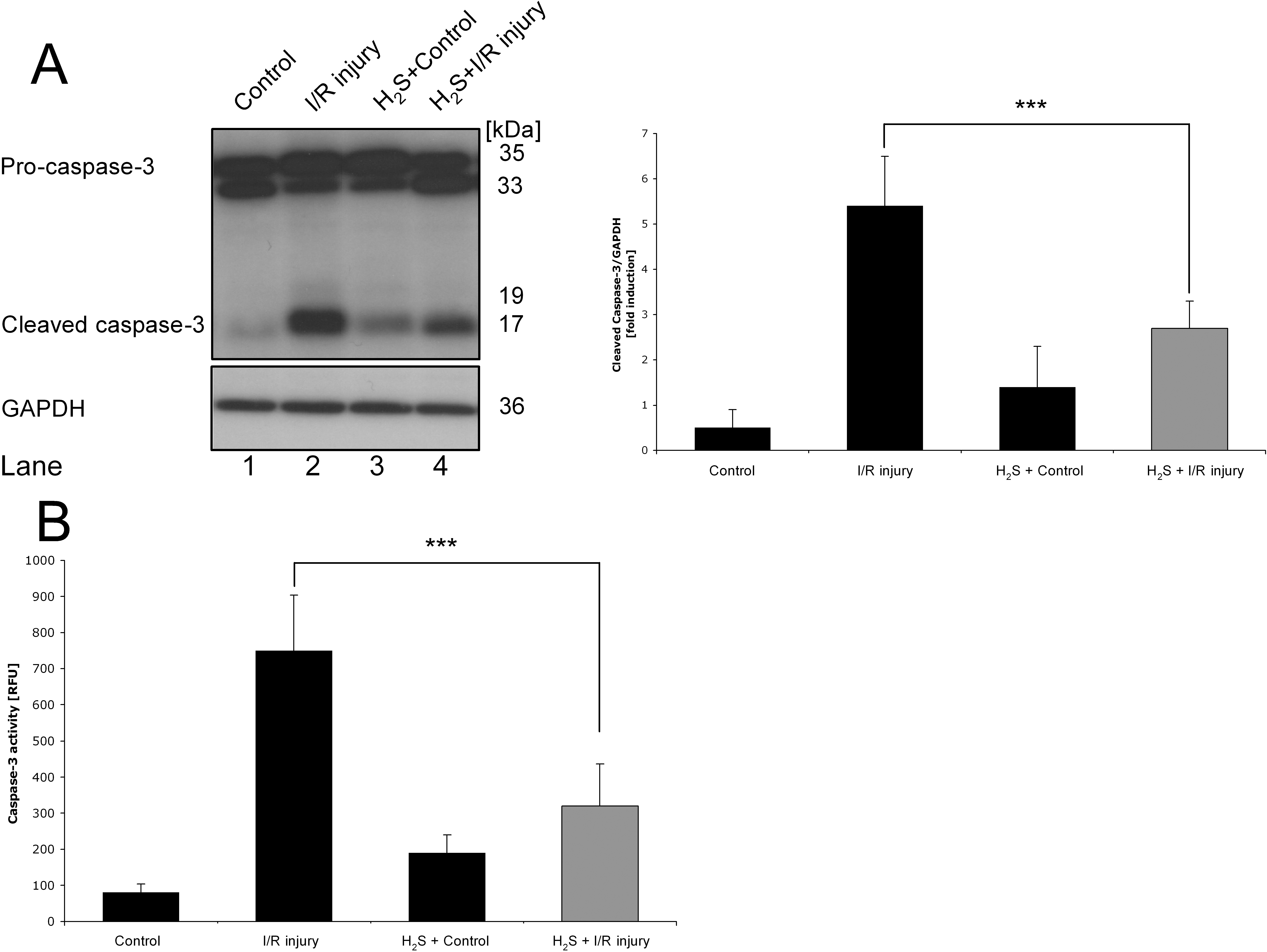
 Figure 2 of Biermann, Mol Vis 2011; 17:1275-1286.