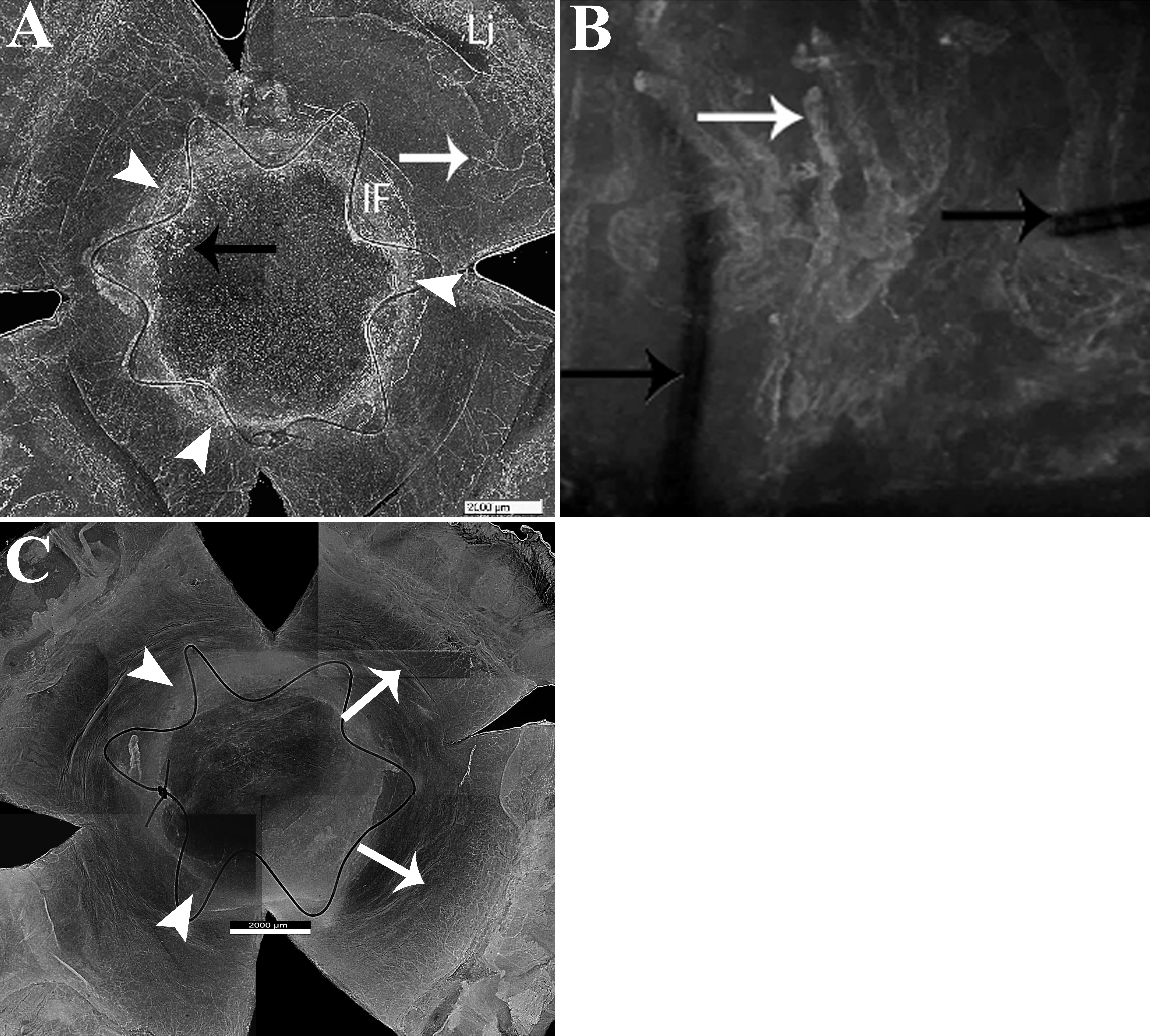
Figure 3. Flat mounted corneas stained for
new vessels 21 days after allotransplantation. Allografted cornea
treated with inactivated (A, B) or active (C) rat
anti-VEGF antibody and flat mounted at Day 21; new vessels
stained with lectin arise from the limbus toward the grafted area (A,
B, C; white arrows). The progression of new
vessels crossed the trephination line (A; arrow heads) across
the stroma of the button (A, black arrow) between the strand of
the running suture (B, black arrow), securing the corneal graft
to the recipient bed. It was limited to the recipient area in treated
animals (C; white arrows). Li, limbus; IF, interface between
donor and recipient (A, C; arrowheads,). Scale Bar=2200
µm. Magnification, A, C 20×, B 100×.

 Figure 3 of Rocher, Mol Vis 2011; 17:104-112.
  Figure 3 of Rocher, Mol Vis 2011; 17:104-112.