Figure 7. Transmission electron
microscopic changes. A: Control, B: Selenite induced, C:
Selenite
+ FVN treated. Lenses were fixed in 2.5% glutaraldehyde and 2%
paraformaldehyde for 12–18 h. Samples were processed, sectioned,
stained and examined under a Hitachi H-600 Transmission electron
microscope. Magnification – 8,400×. Arrows indicates enlarged,
irregularly shaped fibers with opaque material.
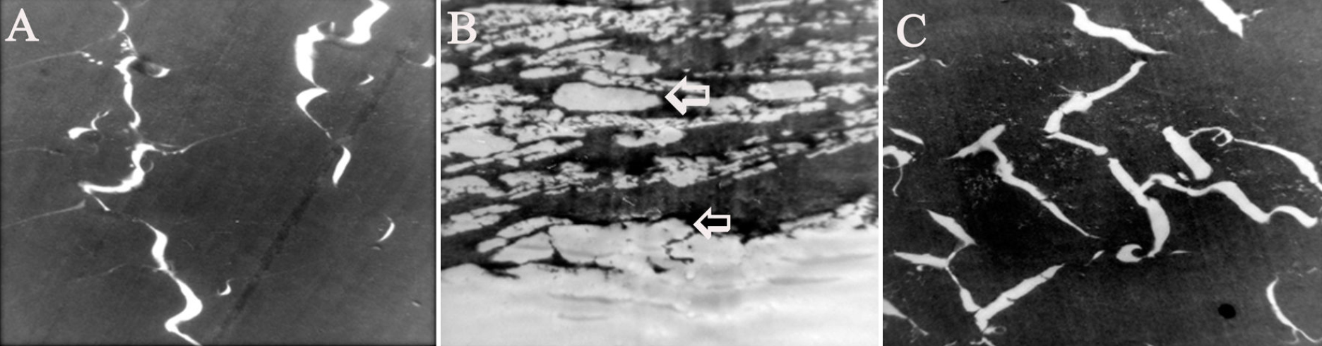
 Figure 7 of Rooban, Mol Vis 2011; 17:1239-1248.