Figure 6. Scanning electron microscopic
changes. A: Control, B: Selenite induced, C:
Selenite + FVN treated. Dissected lenses were fixed in 3%
glutaraldehyde in 0.1 M phosphate buffer at 4 °C. After fixation
the lenses were dehydrated in graded acetone series and critical point
dried. The lenses were then coated with gold and used for the SEM study
under a Philips scanning electron microscope 501(B) at 50 kV.
Magnification – 2,000×. Arrows indicates abnormal fiber structure and
numerous spherical bodies in lens.
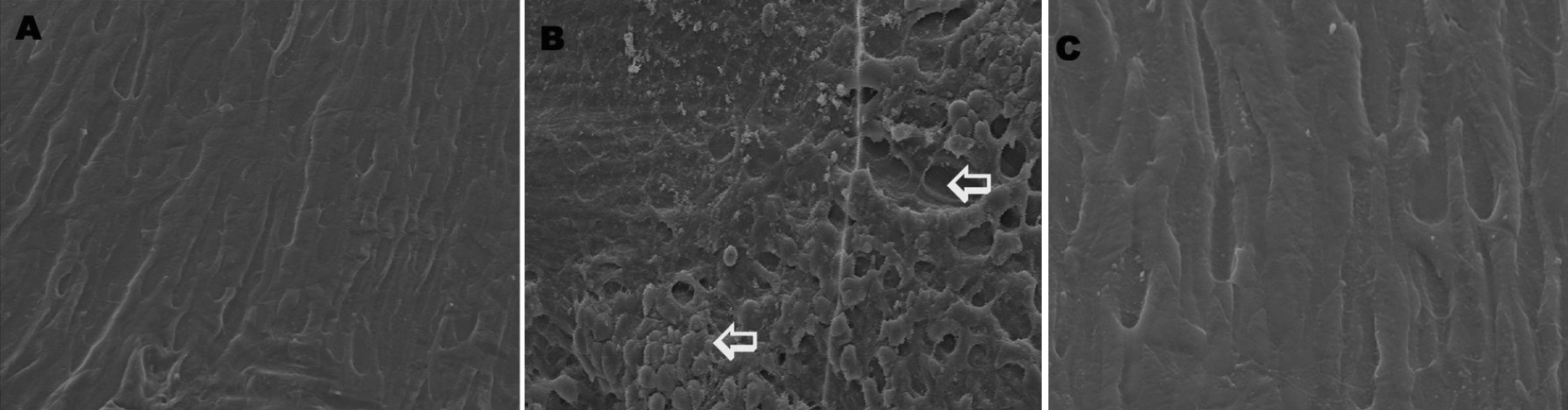
 Figure 6 of Rooban, Mol Vis 2011; 17:1239-1248.