Figure 4. Cellular Localization of Snail in ARPE-19 cells. A: Cellular localization of Snail (red) at 2 days after passage. Snail is observed in both the nuclei and cytoplasm. Nuclei
were counterstained with Hoechst 33258 (blue). B: Cellular localization of Snail (red) at 7 days after passage. Snail expression is decreased and observed mainly in the cytoplasm,
but not in the nuclei. C: Negative control. Scale bar=50 µm.
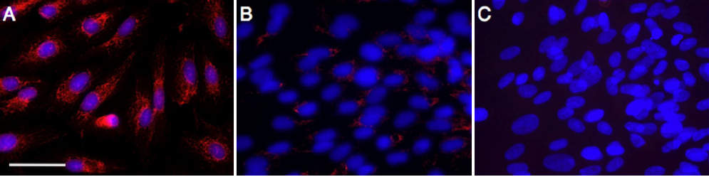
 Figure 4 of
Hirasawa, Mol Vis 2011; 17:1222-1230.
Figure 4 of
Hirasawa, Mol Vis 2011; 17:1222-1230.