Figure 2. Immunolocalization of Snail protein in human CNV. Representative micrographs of human CNV section. A: Phase contrast image. B: Fluorescent micrograph of RPE65 (green). C: Fluorescent micrograph of Snail (red). D: Merged Image. Nuclei were counterstained with Hoechst 33258 (blue). Scale bar=50 µm.
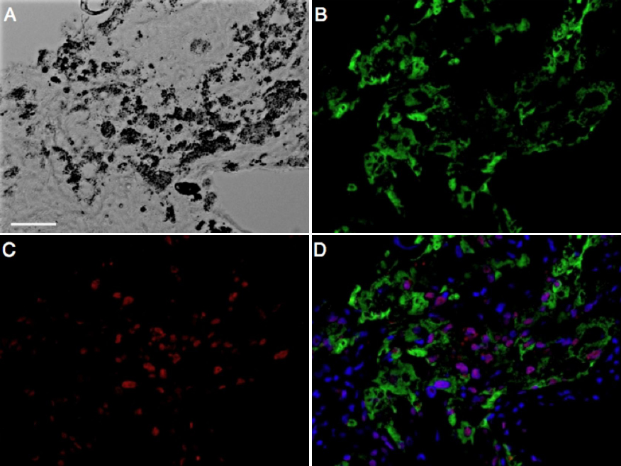
 Figure 2 of
Hirasawa, Mol Vis 2011; 17:1222-1230.
Figure 2 of
Hirasawa, Mol Vis 2011; 17:1222-1230.