Figure 1. Immunolocalization of Snail protein in normal retina. A: Representative immunofluorescent micrograph of Snail (red), RPE65 (green), and nuclear counterstaining with Hoechst 33258
(blue) in normal human retina. B: High magnification. C: Negative control. D: Merged image of a representative immunofluorescent micrograph of Snail (red), RPE65 (green), and nuclear counterstaining
with Hoechst 33258 (blue) in normal mouse retina. Scale bars for A, C, and D shown in A=50 µm, for B=25 µm.
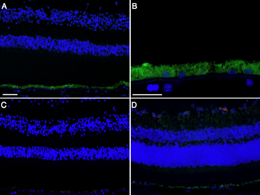
 Figure 1 of
Hirasawa, Mol Vis 2011; 17:1222-1230.
Figure 1 of
Hirasawa, Mol Vis 2011; 17:1222-1230.