Figure 3. Confirmation of effects of PITX2
knockdown on ALDH1A1 and CXCL6 at the protein level.
Results shown are with PITX2 siRNA-1 and TM1. A: Representative
western immunoblots of ALDH1A1 and CXCL6 in protein extracts of TM
cultures exposed to scrambled siRNA and PITX2 siRNA. B-G:
Immunofluorescent
analysis of ALDH1A1expression in TM cultured cells
exposed to scrambled siRNA (B, C) and PITX2
siRNA (D, E). In cells treated with scrambled siRNA (B),
ALDH1A1
expression is apparent in cytoplasm and nucleus of most cells.
However, decreased expression is evident in many PITX2 siRNA
treated cells (D). No immunofluorescence is observed in the
negative control (F). DAPI stained cells are also shown (C,
E, G). H-S: Immunohistochemical
demonstration of ALDH1A1 and CXCL6 expression in the human eye.
Cryosections of anterior section of donated globe observed under light
microscope (H, K), by immunofluoresence after staining
for ALDH1A1 and CXCL6 (I, L, N, O), and
after staining with DAPI (J, M, Q, R)
are shown. H, I, J: sections stained with
anti-ALDH1A1. K, L, M: sections stained with
anti-CXCL6. Expression of both ALDH1A1 and CXCL6 in the trabecular
meshwork (TM) and stroma (S), and higher expression in the Descemet
membrane (DM) are evident. N, Q: highest expression of
ALDH1A1 was observed in the epithelium of the cornea (EP). O, R:
highest
expression of CXCL6 was observed in the iris (I), particularly
in the iris epithelium. P, S: negative control shows no
immunofluorescent staining. Optical lens magnification 5× (all except O
and R), 20× (O and R).
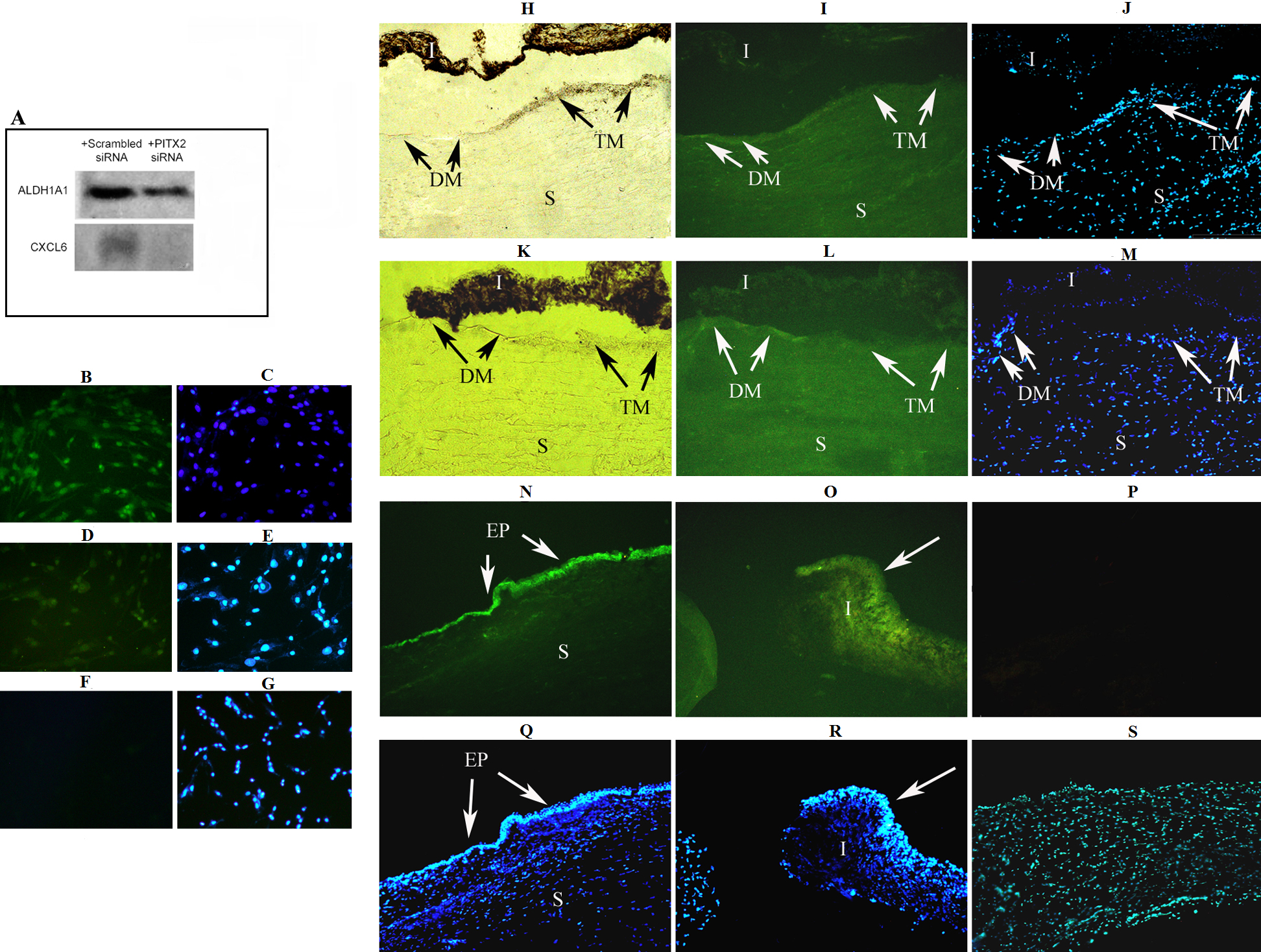
 Figure 3 of Paylakhi, Mol Vis 2011; 17:1209-1221.