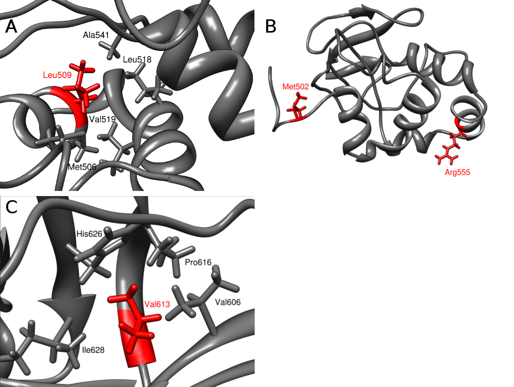
Figure 5. Local structure around the mutated residue in the fourth domain of the keratoepithelin protein (
1x3b PDB code). The protein is shown in cartoon representation. Leu509 (
A), Met502 and Arg555 residues (
B), Val613 (
C) and the neighboring residues are shown in stick representation in red and gray, respectively.
 Figure 5 of
Niel-Butschi, Mol Vis 2011; 17:1192-1202.
Figure 5 of
Niel-Butschi, Mol Vis 2011; 17:1192-1202.