Figure 1. Family A. A: Pedigree showing a four-generation family affected by lattice-type corneal dystrophy. Open and closed symbols indicate unaffected
and affected individuals, respectively, arrows indicate the proband in each family, and asterisks indicate members who were
examined clinically and genetically. A bar on top of a symbol indicates that patients had received a bilateral grafted in
both eyes. Diagonal lines indicate deceased individuals. B: Retroillumination slit-lamp view of patient IV-1 at 21 years of age. The left eye contained a network of thick lattice lines
(arrowhead) and dots. C: Congo red positive deposits of amyloid aggregates are found in subepithelial (fine arrow) and mild corneal stroma (arrowhead).
Green birefringence is visible with a polarizing filter. Stromal deposition of amyloid substance distorts the architecture
of corneal lamellae. D: The electropherogram of exon 11 of the TGFBI gene is shown. The proband III.1 has a heterozygous thymine to cytosine change at codon 509 in exon 11 (c.1526T>C, p.Leu509Pro).
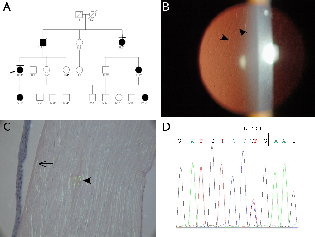
 Figure 1 of
Niel-Butschi, Mol Vis 2011; 17:1192-1202.
Figure 1 of
Niel-Butschi, Mol Vis 2011; 17:1192-1202.