Figure 1. Increased ROS production and diminished anti-oxidant potential in GLC cells. A: Illustration of the levels of malondialdehyde (MDA), a naturally occurring product of lipid peroxidation (an indicator of
oxidative stress) in LC cells. Increased expression of MDA was found in glaucomatous LC cells (GLC) when compared with LC
cells derived from normal donors (NLC; 27.19±7.05 µM MDA versu 14.59 µM MDA±0.82, p≤0.05, n=3). B: mRNA expression of enzymes associated with maintenance of oxidative homeostasis was analyzed. As can be seen, expression
of AKR1C1 and GCLC are significantly lower in GLC cells (p=0.02) compared to NLC cells, expression normalized to NLC gene expression value. These results are suggestive of increased
levels of oxidative stress and/or a compromised anti-oxidant state in cells from glaucomatous donors. Results are representative
of 3 independent experiments with n=3 for NLC and GLC in each experiment.
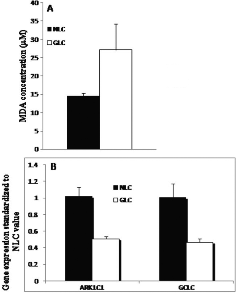
 Figure 1 of
McElnea, Mol Vis 2011; 17:1182-1191.
Figure 1 of
McElnea, Mol Vis 2011; 17:1182-1191.