Figure 4. Histological analysis of Aca47 eyes. Wild-type (WT), heterozygous (A/+), and homozygous mutant eyes are compared at ten weeks of age. A: The overview of the analyzed eye sections indicate that histology of cornea (Co) and retina (Re) is not altered in the mutants.
Le: lens; bars=0.5 mm. B: Magnification of the lens bow regions reveal regular degradation patterns of lens fiber cell nuclei in the mutants. Bars=50
µm. C: Lens fiber layers at the equatorial outer cortex are regularly arranged in wild-types and heterozygous mutants. In homozygous
individuals, fiber layers are partially thickened (arrow) and show less regular, wave-like structures. Bars=20 µm.
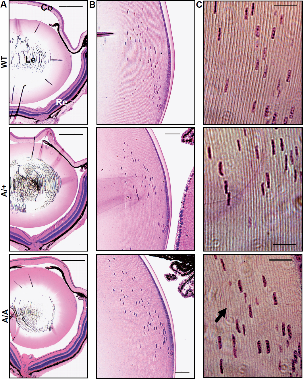
 Figure 4 of
Puk, Mol Vis 2011; 17:1164-1171.
Figure 4 of
Puk, Mol Vis 2011; 17:1164-1171.