Figure 5. Fundus photos of the asymptomatic father with the c.601delC mutation and the unaffected mother without the mutation. A and B: The mother has normal fundi. C-D: Fundus photos of the asymptomatic father shows normal posterior fundi. E and F: The father has increased vessel branching in the equatorial area and a peripheral avascular zone. G and H: Optical coherence tomography scan shows normal macula of the asymptomatic father.
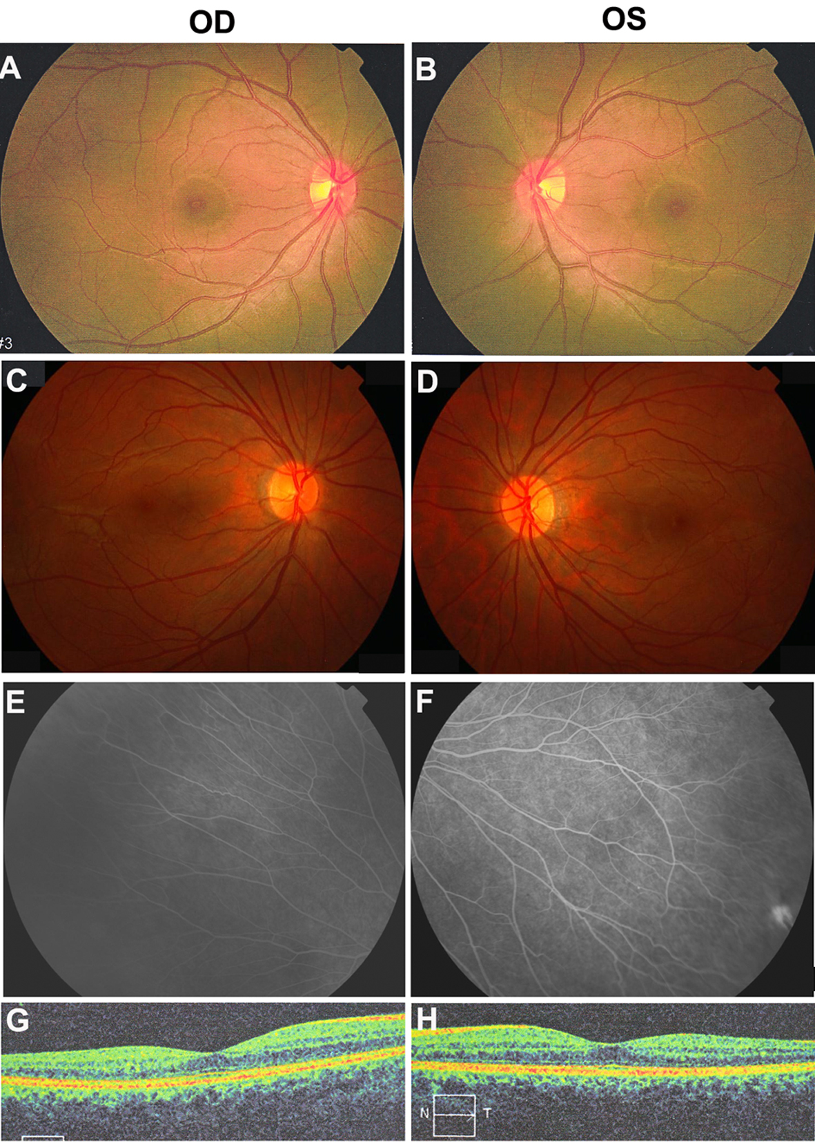
 Figure 5 of
Yang, Mol Vis 2011; 17:1128-1135.
Figure 5 of
Yang, Mol Vis 2011; 17:1128-1135.