Figure 3. Fundus changes in the patient with the heterozygous c.313T>C mutation in Family B. A and B: These color photos demonstrate a normal right posterior fundus and traction of the retinal vessels and macular degeneration
in the left fundus. C and D: Fluorescein angiography of the left eye shows straightening of the vessels with increasing branching (C) and a peripheral avascular zone (D). OD and OS represent the right and left eyes, respectively.
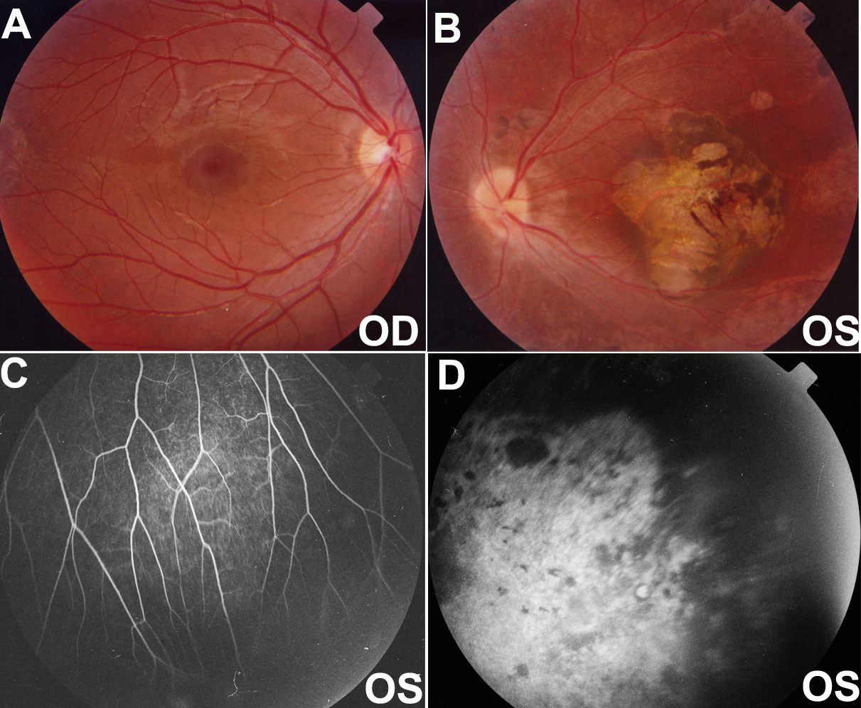
 Figure 3 of
Yang, Mol Vis 2011; 17:1128-1135.
Figure 3 of
Yang, Mol Vis 2011; 17:1128-1135.