Figure 8. Scanning-laser ophthalmoscopy
(SLO) imaging. Eyes from Rosa26 (wt; upper row), Rosa26-floxSTOP/SRF-VP16
(het) (middle row), and Rosa26-floxSTOP/SRF-VP16 (hom; bottom
row) animals at six months of age (P180) were investigated. Fundus
imaging with RF at 514 nm excitation (left column) and autofluorescence
at 488 nm laser wavelengths (middle left column) display dramatic
changes in fundus appearance, structure of the nerve fiber layer and
accumulation of autofluorescent lipids in genetically modified animals
compared to wild-type animals. Retinal degenerative processes can also
be inferred from enhanced angiographic deep layer (choroidal) signals
using fluorescein (middle right column) and indocyanine green (right
column) angiography, as clearly detected in both heterozygous and
homozygous animals.
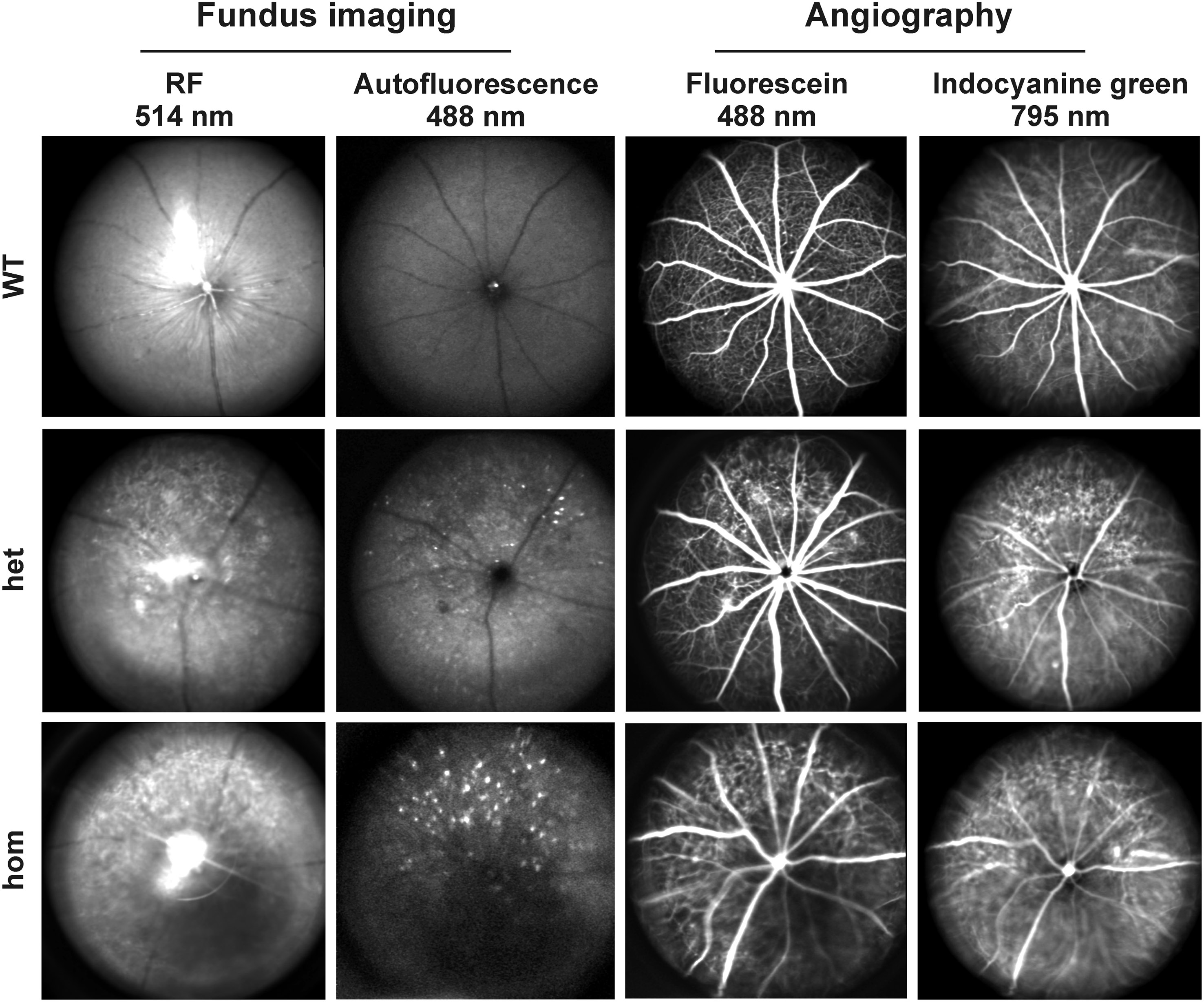
 Figure 8 of Sandström, Mol Vis 2011; 17:1110-1127.