Figure 7. Retinal degeneration in P180
mice heterozygous or homozygous for the Rosa26-floxSTOP/SRF-VP16
transgene. Histological sections of P180 retinas are shown displaying
degenerative processes in Rosa26-floxSTOP/SRF-VP16 heterozygous
(het) and homozygous (hom) animals. A-C: Hematoxylin and
eosin staining. Note the scattered degeneration in the posterior part
of heterozygous and homozygous retinas. Peripheral regions show no
degeneration. Scale bars represent 200 μM. D-F: Higher
magnifications of P180 retinas are displayed. Wild-type retina (D)
showing
ganglion cell layer, inner plexiform layer, inner nuclear
layer, outer nuclear layer, inner and outer segments and retinal
pigment epithelium (RPE). Degeneration of the photoreceptor layer in
adult heterozygous (E) and homozygous (F) retinas is
shown. In (F), arrowheads point to regions of complete
degeneration of the inner and outer nuclear layers, whereas inner
nuclear layers can be found at least partially in other places
(asterisks). Occasionally, even the ganglion cell layer can no longer
be found (indicated by horizontal double-headed arrow). Scale bars
represent 50 μM. G: Quantification of individual mice
displaying retinal degeneration in histological sections. Retinas
displaying areas in the outer nuclear layer with eight or fewer layers
of photoreceptor nuclei were classified as having “partial retinal
degeneration.” Sixty percent of the heterozygous animals and over 80%
of the homozygous animals showed partial degeneration.
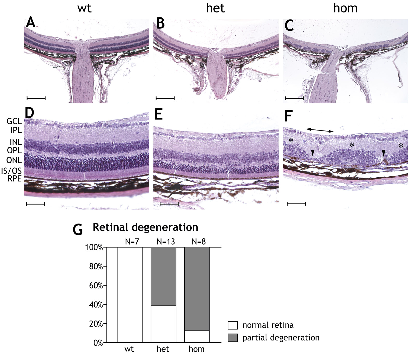
 Figure 7 of Sandström, Mol Vis 2011; 17:1110-1127.