Figure 4. scAAV-mediated mCherry
expression in human retinal pigment epithelial (ARPE19) and 661W cone
photoreceptor cells, at three days post infection. The top row consists
of representative images of mCherry expression in ARPE19 cells infected
with (A) scAAV1, (B) scAAV2, and (C) scAAV2
sextuple mutants at a multiplicity of infection (MOI) of 10,000. The
bottom row consists of representative images of mCherry expression in
661W cells infected with (D) scAAV8, (E) scAAV2, and (F)
scAAV2
sextuple mutants at an MOI of 10,000. All 10× images were taken
with identical exposure times (800 ms). The scale bar in A=200 µm.
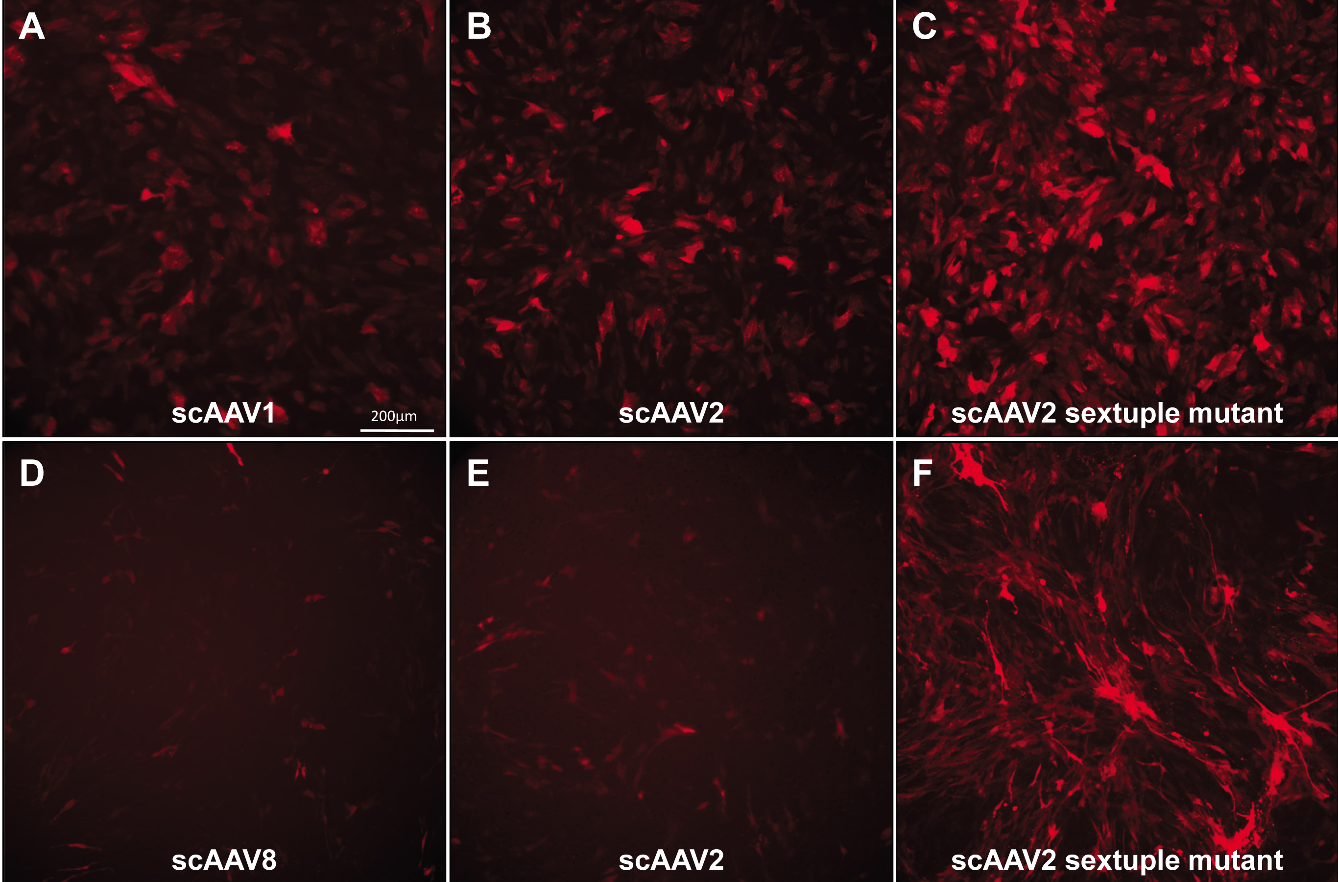
 Figure 4 of Ryals, Mol Vis 2011; 17:1090-1102.