Figure 6. Effect of senescence on antigen
expression and migration of late outgrowth endothelial cells (OECs) and
Human Umbilical Vein Endothelial Cells (HUVEC). A: Expression
of different antigens by control nonsenescent OECs, OECs treated with
SU5416 (6 μM) for 7 days and late-passage naturally senescent OECs was
detected by flow cytometry. The graphs represent the mean±Standard
error of the mean (SEM) percentage of positive cells for OECs derived
from three different patients. * indicates a statistically significant
difference (p<0.05) as compared to control OECs. B:
Migration of control OECs, OECs treated with SU5416 (6 μM) for 7 days
and late-passage naturally senescent OECs to Vascular Endothelial
Growth Factor (VEGF), Stromal Cell derived Factor-1 (SDF-1) and
complete angiogenic medium (EGM-2MV) is shown. The graphs represent the
mean±SEM percentage of migrated cells (as compared to cells migrated to
EBM-2 basal medium without the addition of serum or growth factors) for
OECs derived from three different patients. * indicates a statistically
significant difference (p<0.05) as compared to control OECs.
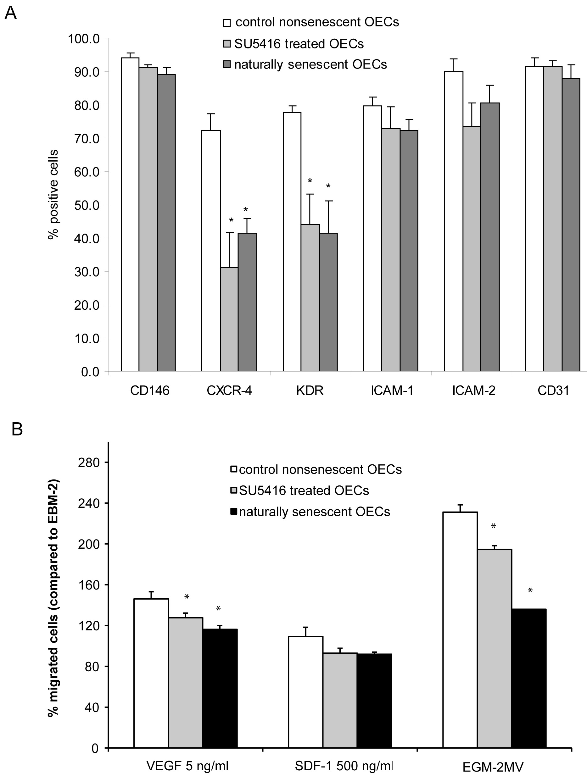
 Figure 6 of Thill, Mol Vis 2011; 17:85-98.