Figure 4. Analysis of telomerase activity
as assayed by the telomeric repeat amplification protocol. A:
Telomerase activity was detected in early passage nonsenescent late
outgrowth endothelial progenitor cells (OECs) and Human Umbilical Vein
Endothelial Cells (HUVEC) cultured in complete angiogenic medium
(EGM-2MV). Negative control represents telomerase activity of
heat-inactivated OEC samples, positive control corresponds to the tumor
cell sample provided by the manufacturer (TeloTAGGG Telomerase PCR
ELISA, Roche Applied Science, Indianapolis, IN). NS denotes not
statistically significant difference in telomerase activity between
HUVEC and OECs. B: Telomerase activity is decreased in OECs
treated with 3, 6 and 10 μM SU5416 for 3 days compared to control
EGM-2MV (with the addition of 10 μl DMSO/ml, 0 μM SU5416). C:
Telomerase activity is decreased in OECs treated with EGM-2MV
supplemented with inhibitors SU5416 (6 μM), ZM323881 (10 μM), KRN633 (4
μM), Wortmannin (100 nM), Ly 294002 (5 μM) and Bisindolylmaleimide I (1
μM) for 3 days compraed to control medium (EGM-2MV with the addition of
10 μl DMSO/ml). D: Recovery of telomerase activity is
dose-dependent. Telomerase activity was assessed for control OECs at
day 0 and 7, OECs inhibited for 7 days with SU5416 3 μM and 6 μM and
for OECs returned to EGM-2MV without inhibitor for another 3 days after
7 days of inhibition. Graphs A and C represent the
mean±SEM telomerase activity, B and D the mean
percentage in telomerase activity as compared to control (medium
without inhibitor in B, medium without inhibitor after 10 days
of culture in D), for three independent experiments each (OECs
derived from three different patients). Student’s t-test for
paired data was used for statistical comparison between control and
inhibitory conditions. * indicates a statistically significant
difference (p<0.05) as compared to control.
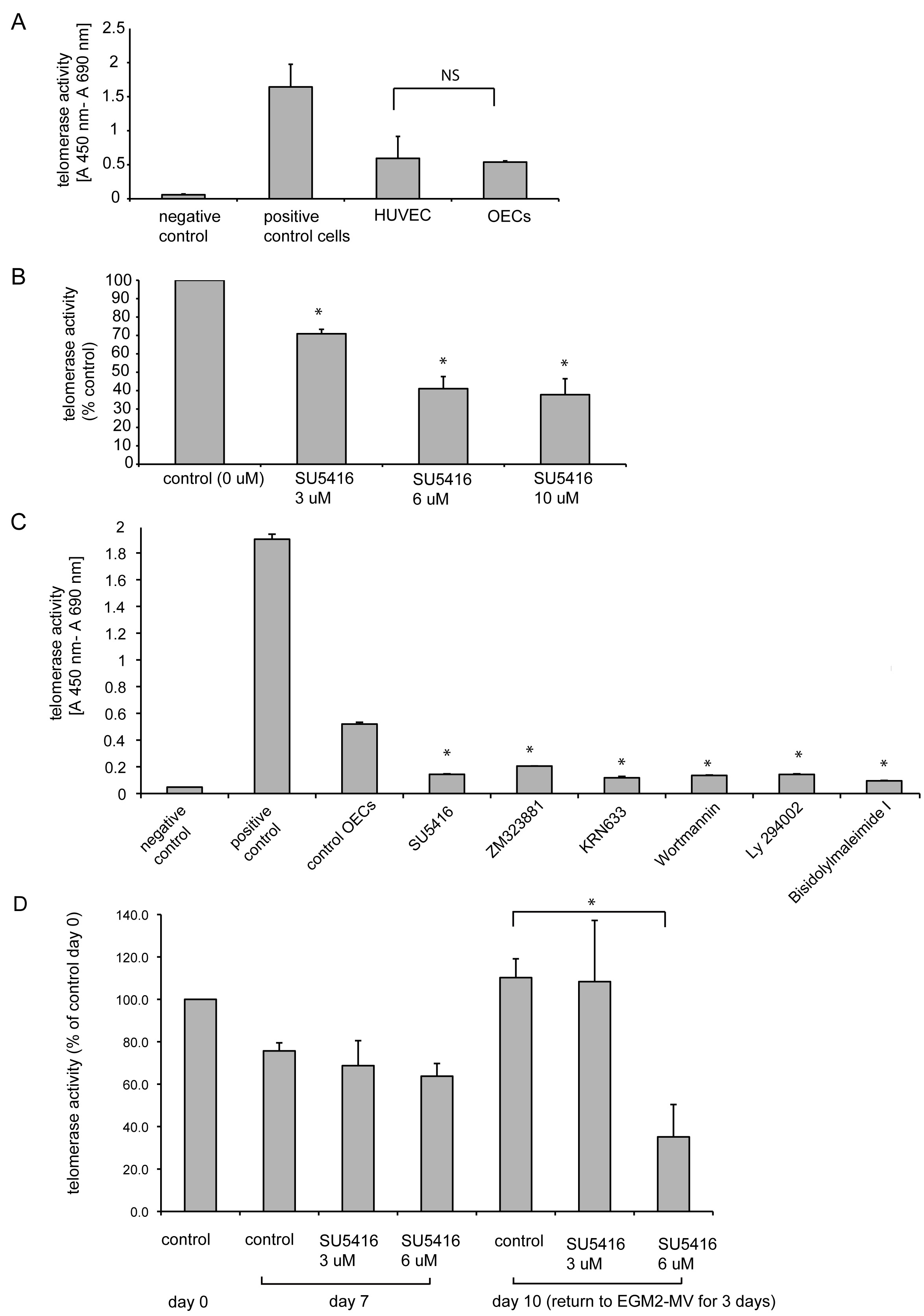
 Figure 4 of Thill, Mol Vis 2011; 17:85-98.