Figure 3. Representative images of
morphology of early passage control late outgrowth endothelial
progenitor cells in complete angiogenic medium containing dimethyl
sulfoxide (10 μl/ml; A), late passage OECs (B), control
early passage OECs after 3 days of inhibition with 10 μM SU5416 (C),
and
3 days after returning SU5416 treated cells to EGM-2MV without
inhibitor (D) are shown. Representative images of
senescence-associated beta-galactosidase staining (blue color) in early
passage control OECs (E), late passage OECs (F) and
early-passage OECs treated with either 10 uM SU5416 (G), 100 nM
Wortmannin (H) or 1 uM Bisindolylmaleimide I (I) for 3
days are shown.
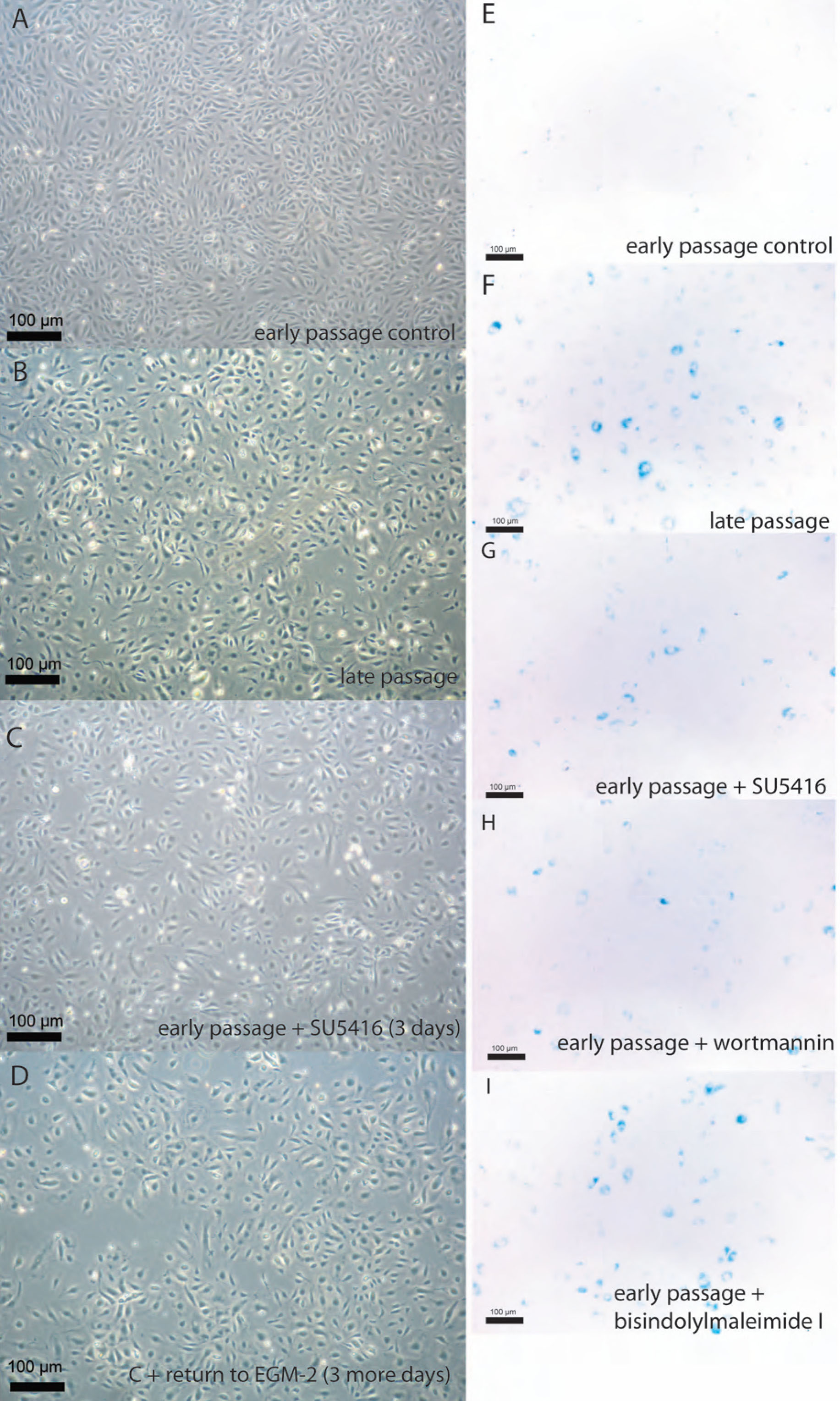
 Figure 3 of Thill, Mol Vis 2011; 17:85-98.