Figure 1. Effect of 48 h of inhibition of
outgrowth endothelial progenitor cells and human umbilical vein
endothelial cells on percentage of apoptotic cells, as determined by
flow cytometric analysis of Annexin V fluorescein isothiocyanate
positive/propidium iodide (PI) negative cells. Cells were grown in
complete angiogenic medium (EGM-2MV) control conditions (10 μl DMSO/ml
EGM-2MV), or in the presence of 1, 3, and 10 μM SU5416, 4 μM KRN633,
and inhibitors of the downstream mediators Akt (100 nM Wortmannin), PI
3-kinase (5 μM Ly 294002), and PKC (1 μM bisindolylmaleimide I). The
graphs represent the mean±SEM cell number from three independent (OECs
were derived from three different patients, for HUVEC analysis was
repeated at least twice) experiments. Student’s t-test for
paired data was used for statistical comparison between control and
inhibitory conditions. P values <0.05 were considered significant
and are denoted by an asterisk in the figure.
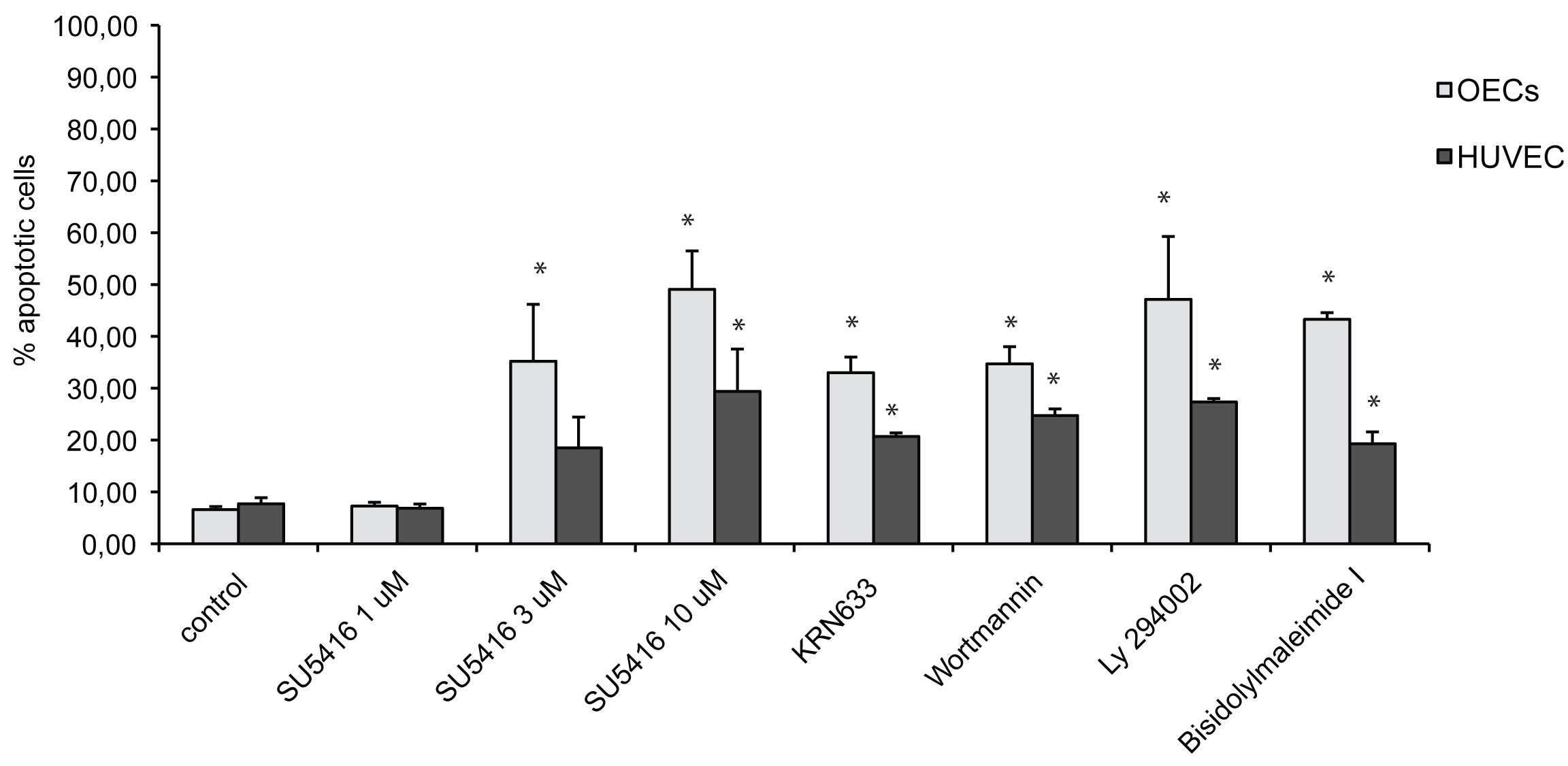
 Figure 1 of Thill, Mol Vis 2011; 17:85-98.