Figure 7. Immunochemistry of apelin in a
retina. Sections of retina were examined by immunohistochemistry with
anti-apelin antibody. Positive staining (brown) of apelin was detected
in the retinal vessel walls, inner nuclear layer, and outer nuclear
layer in the central retinal vein occlusion groups (A: group “1
w,” B: group “2 w,” C: group “24 w”). Among the
intravitreal bevacizumab (IVB) groups (D: group “1 w+IVB,” E:
group
“2 w+IVB,” F: group “24 w+IVB”), only “24 w+IVB” group
had positive staining of apelin in nuclear layers.
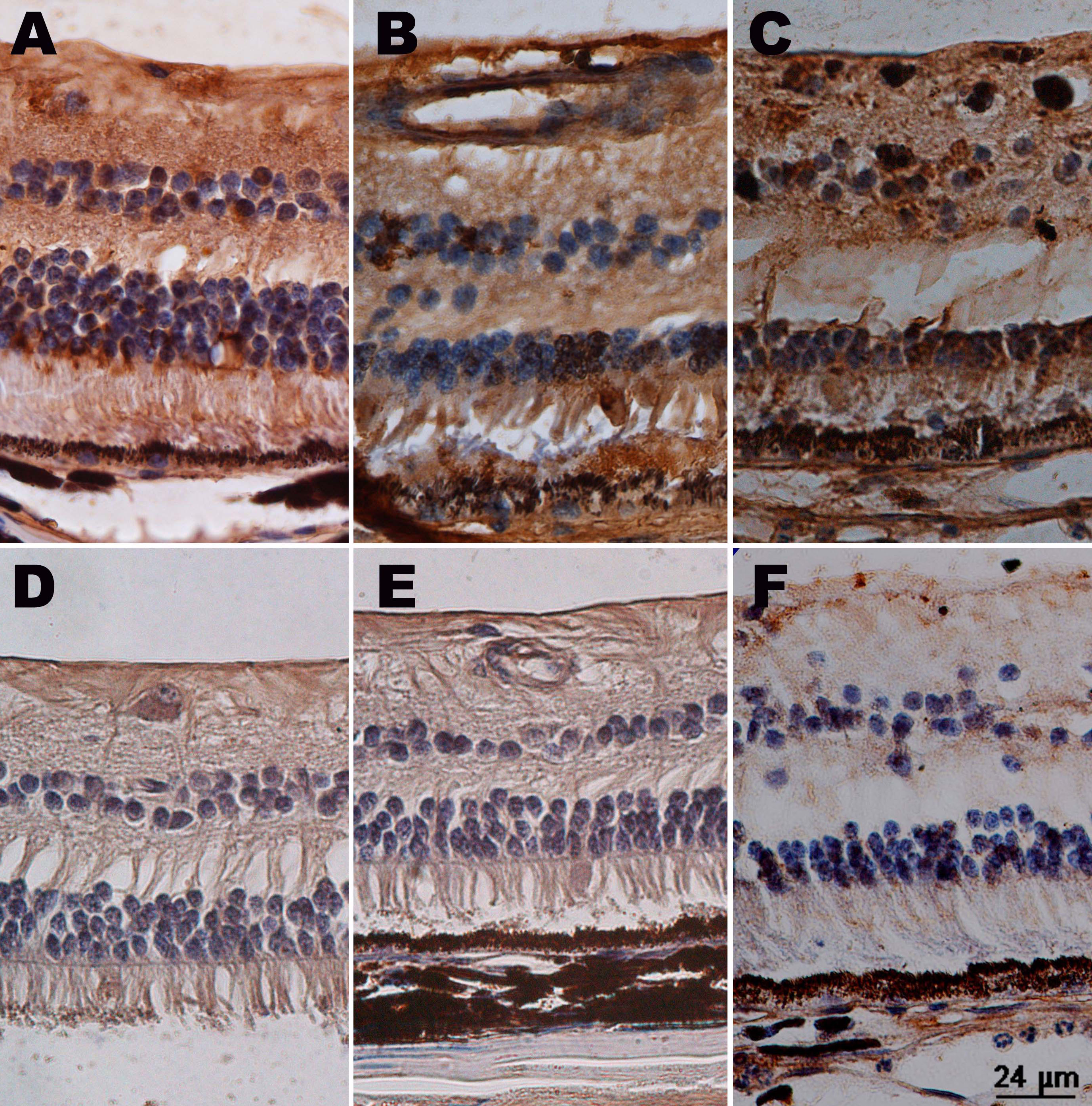
 Figure 7 of Zhao, Mol Vis 2011; 17:1044-1055.