Figure 6. Immunochemistry of vascular
endothelial growth factor in an iris. Sections of iris were examined by
immunohistochemistry with anti-vascular endothelial growth factor
(VEGF) antibody. Positive staining (brown) of VEGF was detected in
vessel walls of iris in the central retinal vein occlusion groups (A:
group
“1 w,” B: group “2 w,” C: group “24 w”). There
was no obvious positive staining after intravitreal bevacizumab (IVB)
injection (D: group “1 w+IVB,” E: group “2 w+IVB,” F:
group
“24 w+IVB”).
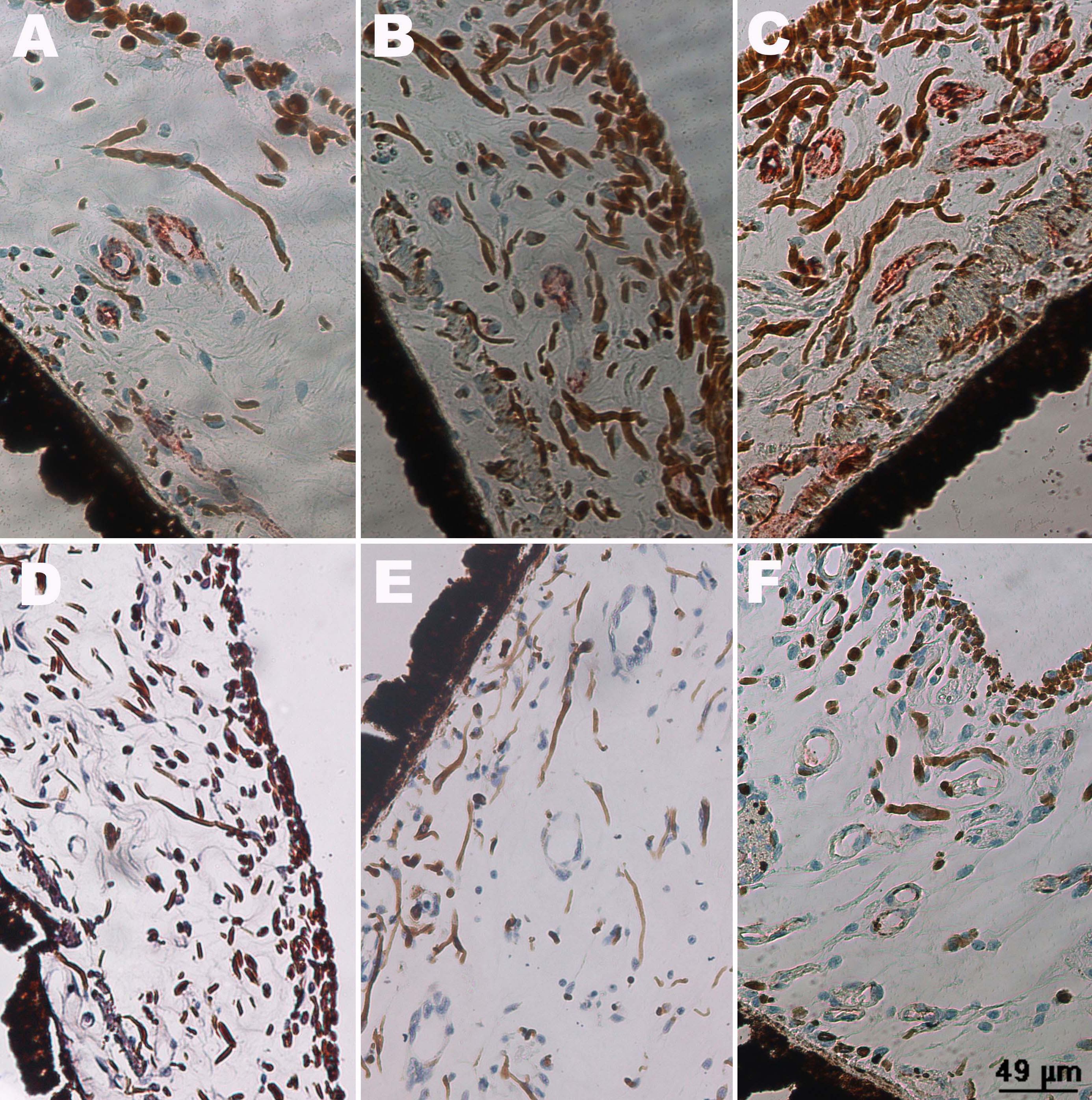
 Figure 6 of Zhao, Mol Vis 2011; 17:1044-1055.