Figure 3. Fluorescence immunostaining of eye tissues after inravitreal bevacizumab (IVB) injection. 4’,6’-diamino-2-phenylindole (blue)
was used to show nuclei and positive staining of bevacizumab (green) was showed in blood vessels. A: Bevacizumab (green) was detected in choroid vessels four weeks after IVB. B: Bevacizumab (green) was detected in the walls of iris vessel four weeks after IVB.
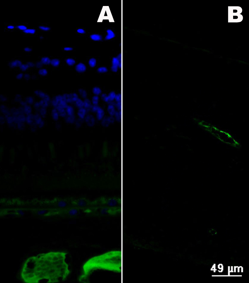
 Figure 3 of
Zhao, Mol Vis 2011; 17:1044-1055.
Figure 3 of
Zhao, Mol Vis 2011; 17:1044-1055.