Figure 2. Hematoxylin and eosin staining
section. With the establishment of central retinal vein occlusion
(CRVO) model, the pathological changes of retina were remarkable. A:
This
image shows the normal retina structure. B: This image
shows interstitial edema of the retina 7 days after photocoagulation. C:
This
image shows disordered retinal structure 24 weeks after
photocoagulation.
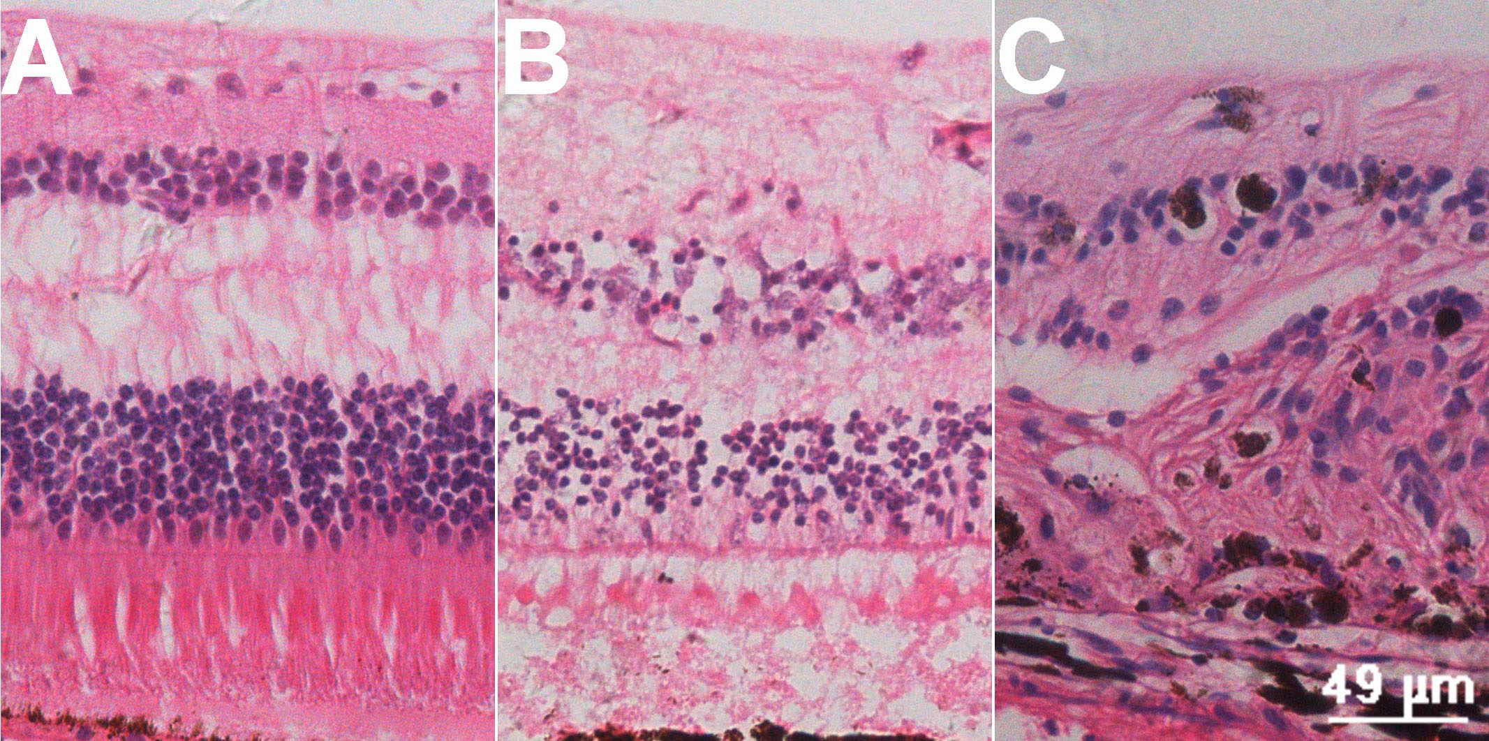
 Figure 2 of Zhao, Mol Vis 2011; 17:1044-1055.