Figure 1. Photographs of fundus. A:
This
shows the normal fundus. B: Central retinal vein occlusion
(CRVO) model was successfully established by photocoagulation. The
fundus photograph shows photocoagulation spots and dilated retinal
veins at the first day after photocoagulation.
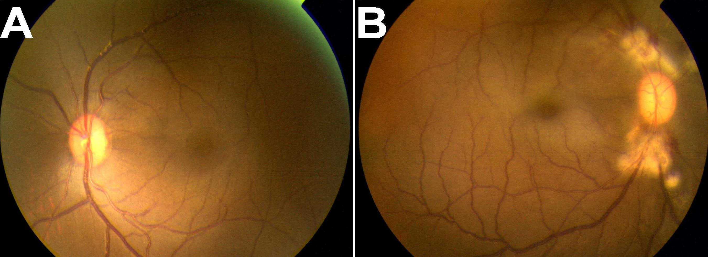
 Figure 1 of Zhao, Mol Vis 2011; 17:1044-1055.