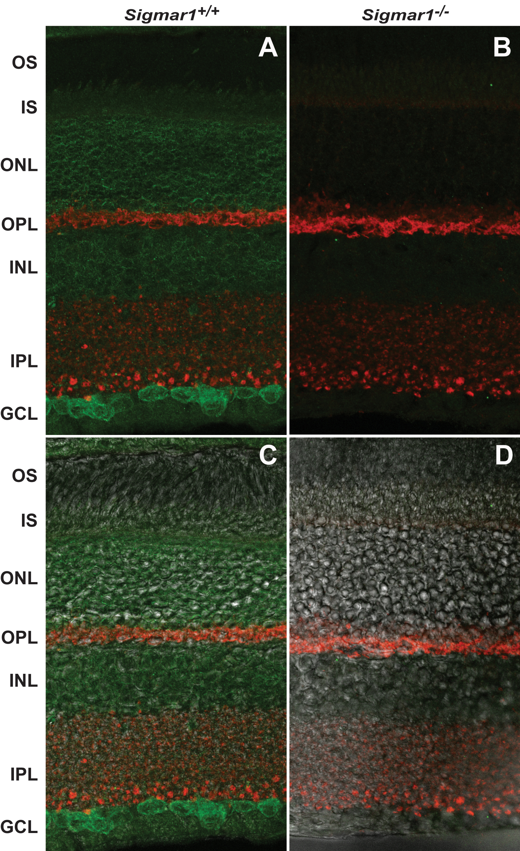
Figure 2. The sigma-1 receptor
distribution in the mouse retina.
A: Immunostaining of the σR1
(green) in the WT mouse retina.
B: Immunostaining of the σR1 in
the
Sigmar1−/− mouse retina (negative control).
C
and
D: The Nomarsky image superimposed with the staining images
in
A and
B, respectively. The abbreviations of the
distinct cell layers are: OS, outer segment; IS, inner segment; ONL,
outer nuclear layer; OPL, outer plexiform layer; INL, inner nuclear
layer, IPL, inner plexiform layer; GCL, ganglion cell layer.
Synaptophysin was stained (red) to mark the presynaptic terminals in
the OPL and IPL. Immunostaining of the σR1 was performed on the mouse
retinal cryosections using the antibody raised against the full-length
σR1 [
6,
32], followed by
incubation with the Alexa-488 conjugated goat-antirabbit antibody. WT
and
Sigmar1−/− mice of the same age (3 months) were
used for preparation of retinal sections. Images were taken on a Nikon
A1R laser confocal microscope and processed using Adobe Photoshop.
 Figure 2 of Mavlyutov, Mol Vis 2011; 17:1034-1043.