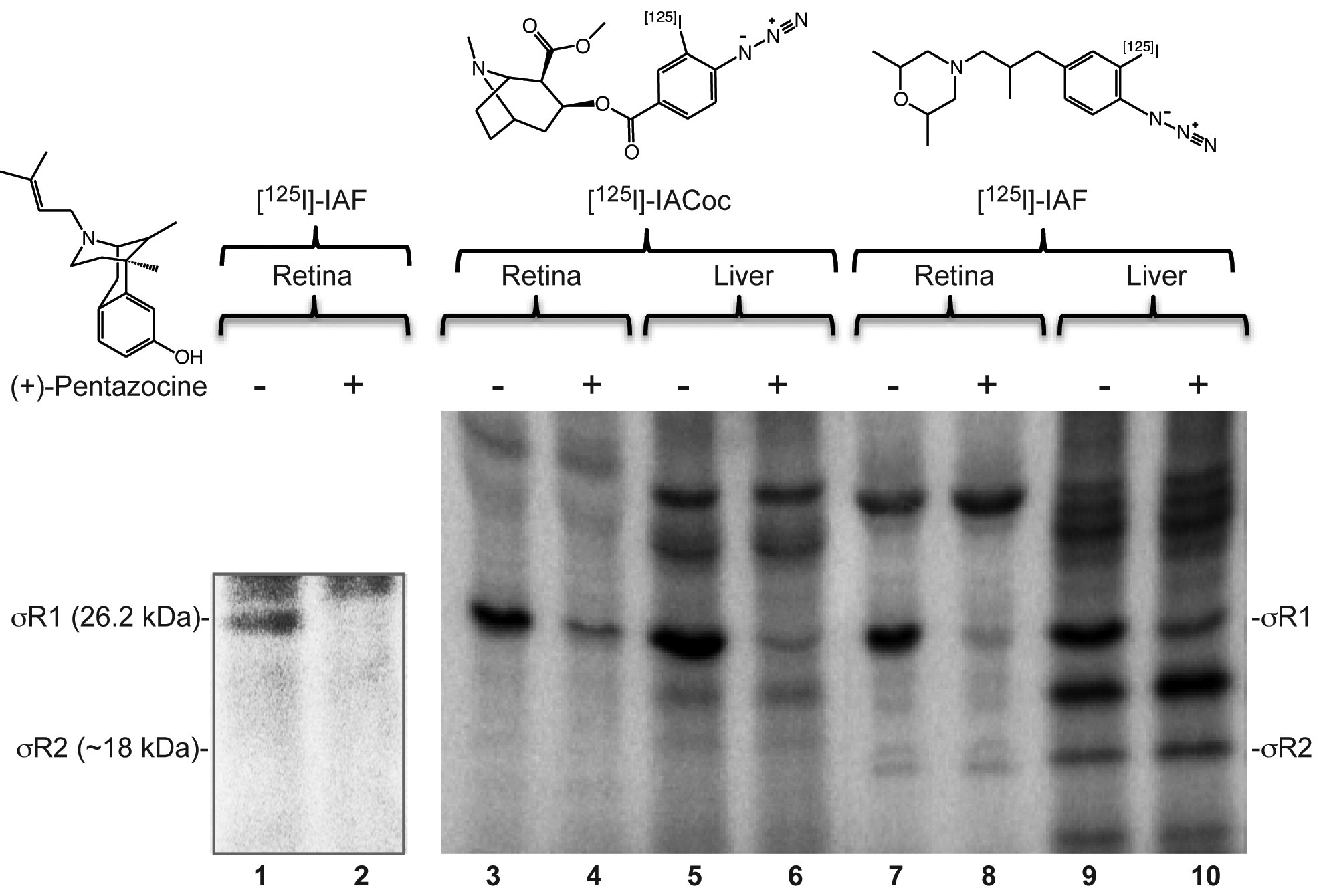
Figure 1. Photoaffinity labeling of the
sigma-1 receptor in the retina. Shown are autoradiograms of the
photolabeled retinal homogenates that were resolved on sodium dodecyl
sulfate gels. Photoaffinity labeling of σRs in the mouse retinal
homogenates was performed with [
125I]-IAF (lanes 1 and 2).
In bovine retinal homogenates, [
125I]-IACoc (lanes 3 and 4)
and [1
25I]-IAF (lanes 7 and 8) were used, and labeling was
compared to that in rat liver membranes (lanes 5 and 6, 9 and 10,
respectively). The specificity of the σR1 photolabeling was
demonstrated by the reduced intensity in the presence of the σR1 ligand
(+)-pentazocine (the even-numbered lanes). In contrast, the
nonspecifically labeled upper bands were not affected by
(+)-pentazocine. The typical labeling pattern of the σR1/σR2 bands
(seen in lane 9) have often been observed previously in mouse and rat
liver membranes [
23,
30]. In each lane of
the gel, 200 μg of total protein was loaded.
 Figure 1 of Mavlyutov, Mol Vis 2011; 17:1034-1043.