Figure 3 of
Yiğit, Mol Vis 2011; 17:1024-1033.
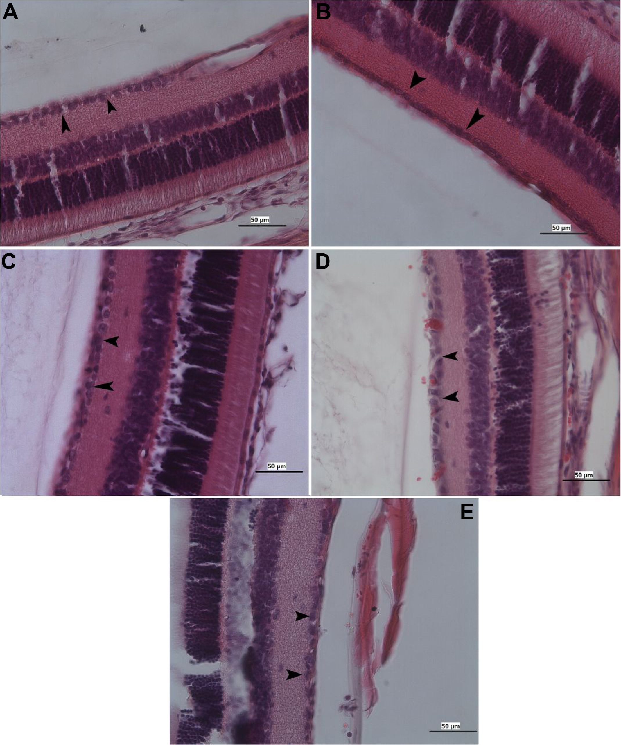
Figure 3.
Photomicrographs showing the ganglion cell (black arrow) of the retina from the mature rat.
A
: control,
B
: ARI,
C
: memantine,
D
: HBO therapy, and
E
: brimonidine. Hematoxylin and eosin stain; Bar 50 µm.
 Figure 3 of Yiğit, Mol Vis 2011; 17:1024-1033.