Figure 2. The effect of raised intraocular
pressure-induced ischemia for 60 min and reperfusion for 21 days on the
percentage of apoptotic (TUNEL positive) cell in rat retina. There is a
significant decrease of apoptotic (TUNEL positive) cell (+) cells
(***p<0.001) in the control retina compared to treatment groups
(MEM, HBO, and BRI) in the left-side graphs. However, there is a
significant increase of apoptotic cells (***p<0.001) in retinas
subjected to ischemia/reperfusion compared to treatment groups in the
right-side graphs. Error bars are ±SEM, where n=6.
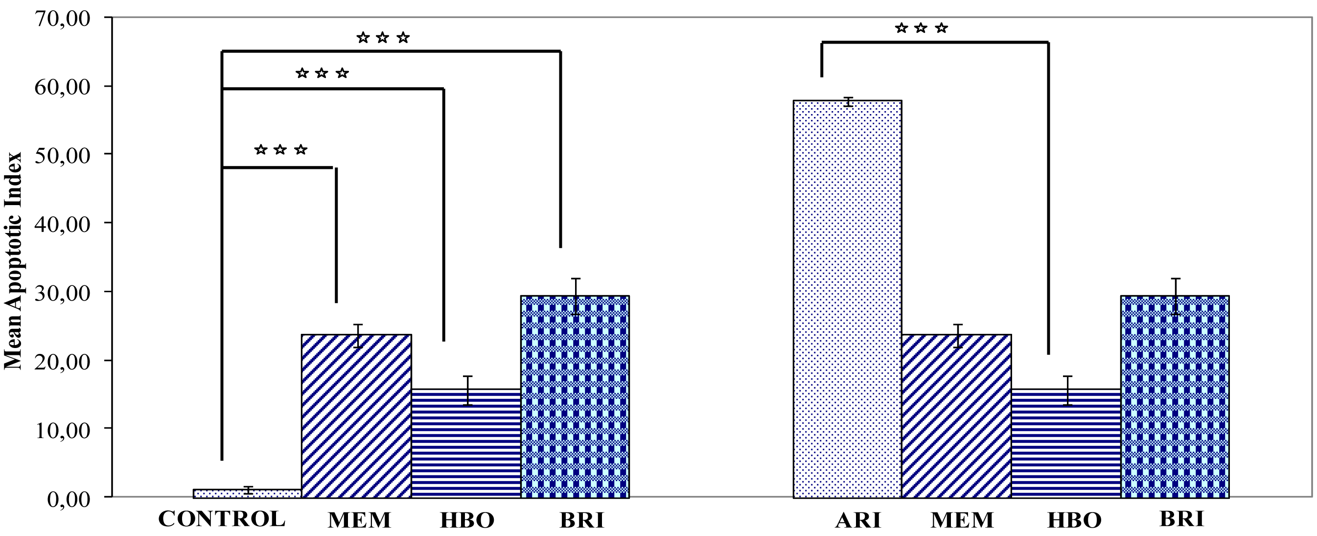
 Figure 2 of Yiğit, Mol Vis 2011; 17:1024-1033.