Figure 2. The percentage of apoptotic
cells is lower in the two strains with hypopigmented retinal pigment
epithelium. The micrographs of the upper panel show sagittal sections
of embryonic (E) day 10.5 (A),
E11.5 (B), and E12.5 (C) BALB/c optic cups and those of
the lower panel show sagittal sections of E10.5 (D), E11.5 (E), E12.5 (F) pJ optic cups after a terminal
deoxynucleotidyl transferase dUTP nick end labeling (TUNEL) assay. RPE,
retinal pigment
epithelium; NR, neuroretina. Scale bars represent 100 μm. Arrows
indicate labeled cells.
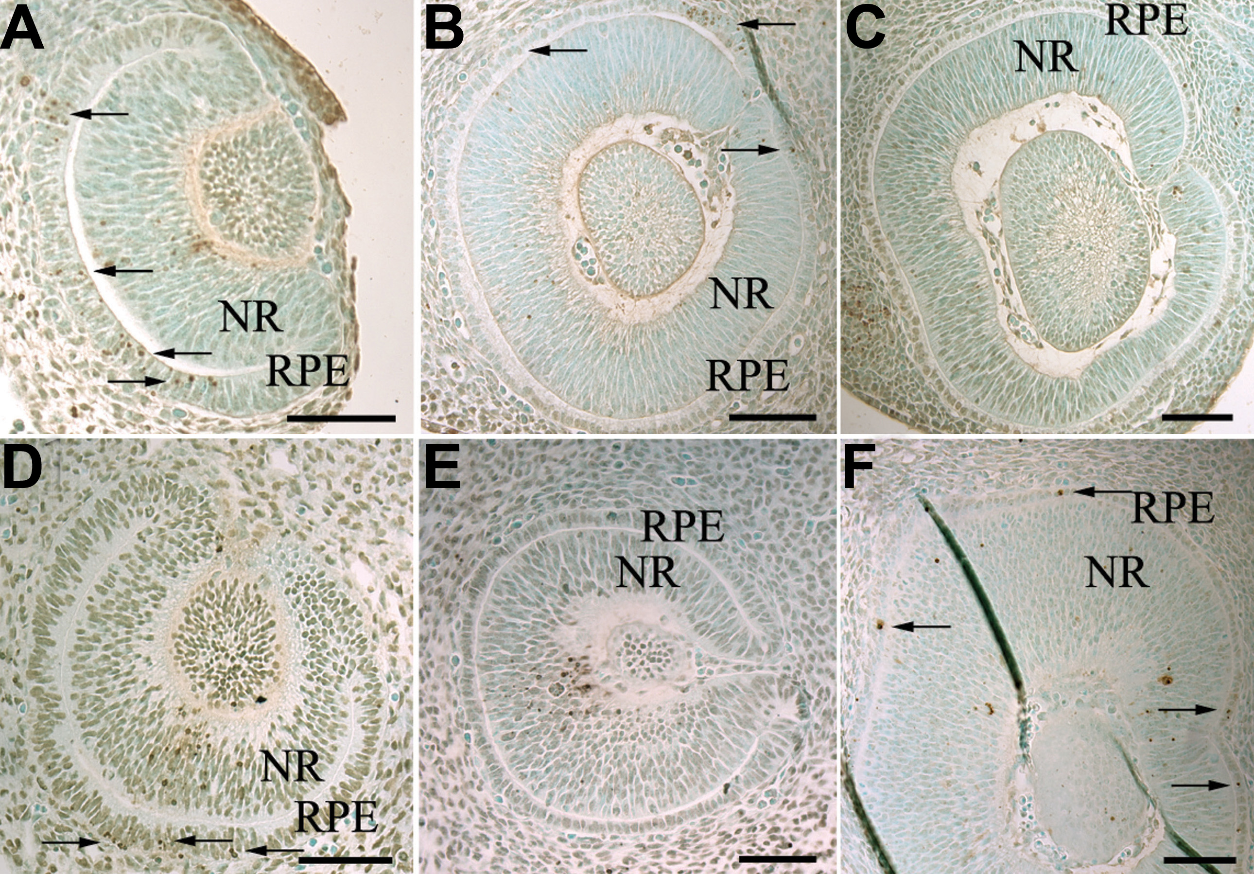
 Figure 2 of Pequignot, Mol Vis 2011; 17:989-996.