Figure 1. Apoptosis is massive in the B6 retinal pigment epithelium at E10.5. A: On sagittal sections of embryonic day (E) 12.5 (left and middle panel) and E11.5 (right panel), B6 RPE is shown at the same
magnification used for counting. We can easily differentiate melanosomes (small black dots) and terminal deoxynucleotidyl
transferase dUTP nick end labeling (TUNEL)-positive cells (brown-labeled dots, indicated by arrows). B: Micrographs in B show sagittal sections of E10.5, E11.5, and E12.5 B6 optic cups after a TUNEL assay. The insets in E10.5 and E11.5 show higher
magnification of apoptotic cells at the corresponding stages. Panel C shows micrographs of frontal sections of E10.5, E11.5, and E12.5 B6 optic cups after a TUNEL assay. RPE, retinal pigment
epithelium; NR, neuroretina; Scale bars represent 100 μm. Arrows indicate examples of RPE-labeled cells (apoptotic cells can
also be observed in the neuroretina), which are predominantly located in the ventral nonpigmented part.
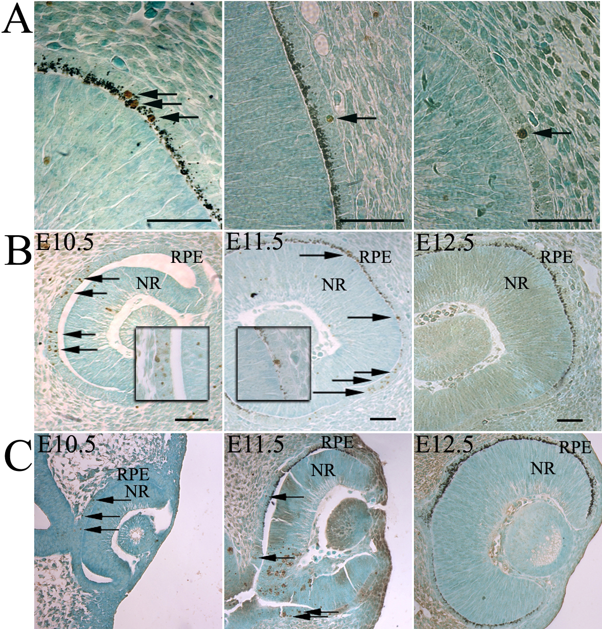
 Figure 1 of
Pequignot, Mol Vis 2011; 17:989-996.
Figure 1 of
Pequignot, Mol Vis 2011; 17:989-996.