Figure 2. Results of sequencing analysis,
PCR analysis, and single-base primer extension assay. A:
Nucleotide and amino acid sequence of TACSTD2. Arrowheads indicate the
site of the c.675C>A and c.840_841insTCATCATCGCCGGCCTCATC nucleotide
changes. Note that the amino acids in bold italic type are of the
transmembrane domain. B: Computationally-predicted domain
structure of the TACSTD2 protein with mutations of previous reports and
this report (*). SS: signal sequence; EGF: EGF-like repeat; TY:
thyroglobulin type I repeat; TM: transmembrane domain; PIP2: PIP2
binding sequence. C: Results of sequencing analysis of TACSTD2
in a normal volunteer (upper) and in proband A or B (lower). The
underlined nucleotides indicate the inserted 20-base sequence between
the 840th and 841st nucleotide positions of TACSTD2. Note that
the presented sequence is in a reverse direction. D: Results of
sequencing analysis of TACSTD2 in a normal volunteer (upper)
and in proband C (lower). Arrowheads indicate the site of the
c.675C>A mutation. Note that the presented sequence is in a reverse
direction. E: Results of PCR analysis to examine the difference
in length between the normal and insertion-bearing alleles in the
families of the proband A (left) and proband B (right). The upper bands
indicate the PCR product derived from the insertion-bearing alleles
while the lower bands indicate the PCR product from the normal alleles.
Note that the sister of proband A was not examined. F: Results
of 1-base primer extension analysis for the 675th nucleotide of TACSTD2
in the normal volunteer (left) and the proband C (right). Black
indicates C and green indicates A. Note that the presented data was
produced by the forward primer.
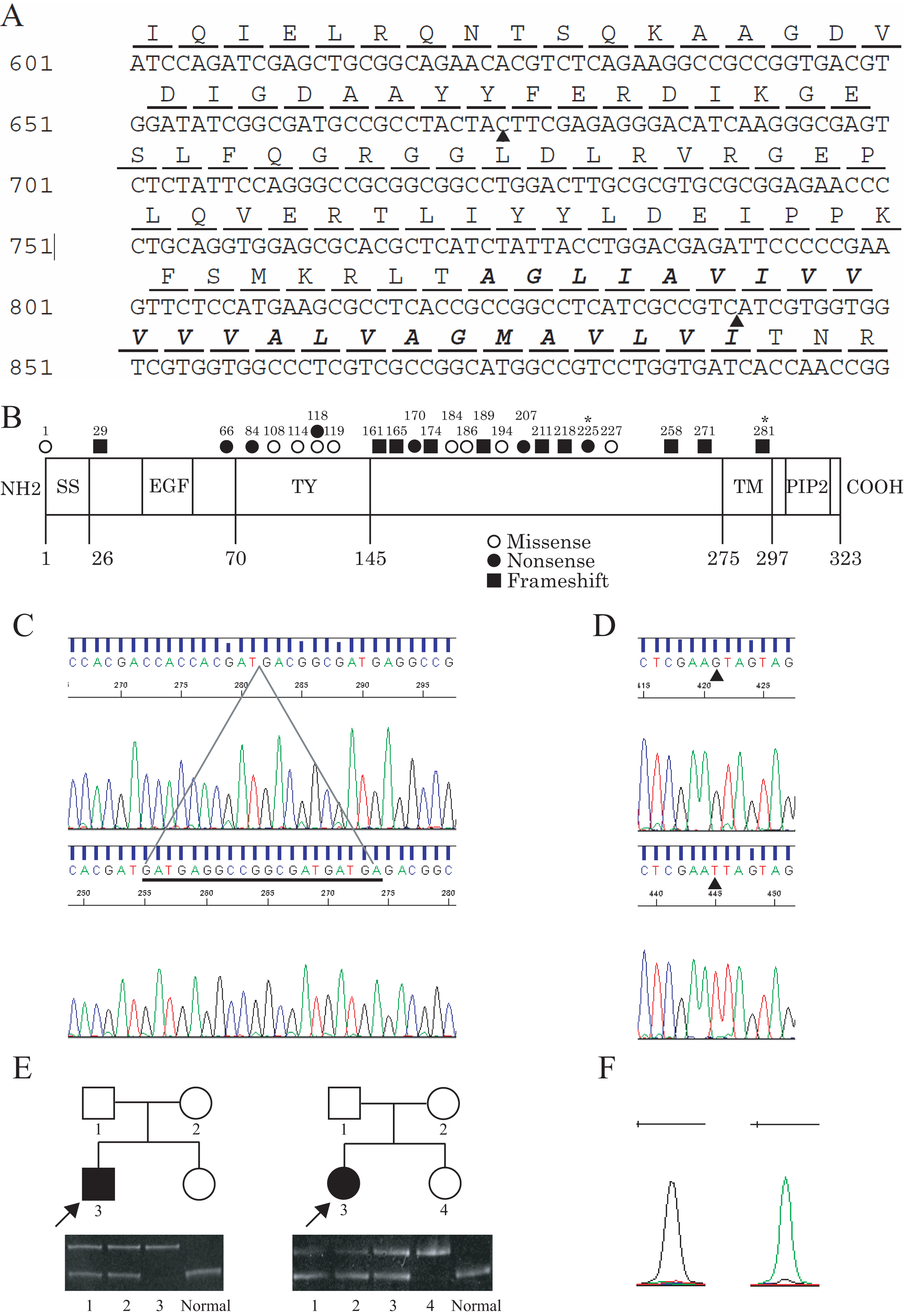
 Figure 2 of Nakatsukasa, Mol Vis 2011; 17:965-970.