Figure 1. Representative anterior segment
photographs from an AKC patient before and after treatment with
tacrolimus ophthalmic solution. Anterior segment photograph (right)
shows an extensive corneal damage visualized by the fluorescein
staining. Note that the superficial punctate keratopathy is present in
almost all the surface of the cornea and is associated to the increased
papillary formation. The photographs on the left side represent the
cornea and papillary formation from the same patient after 1 month of
treatment with 0.1% tacrolimus ophthalmic solution. Note the
improvement of the corneal damage as well as the decrease of papillary
formation and conjunctival inflammatory status.
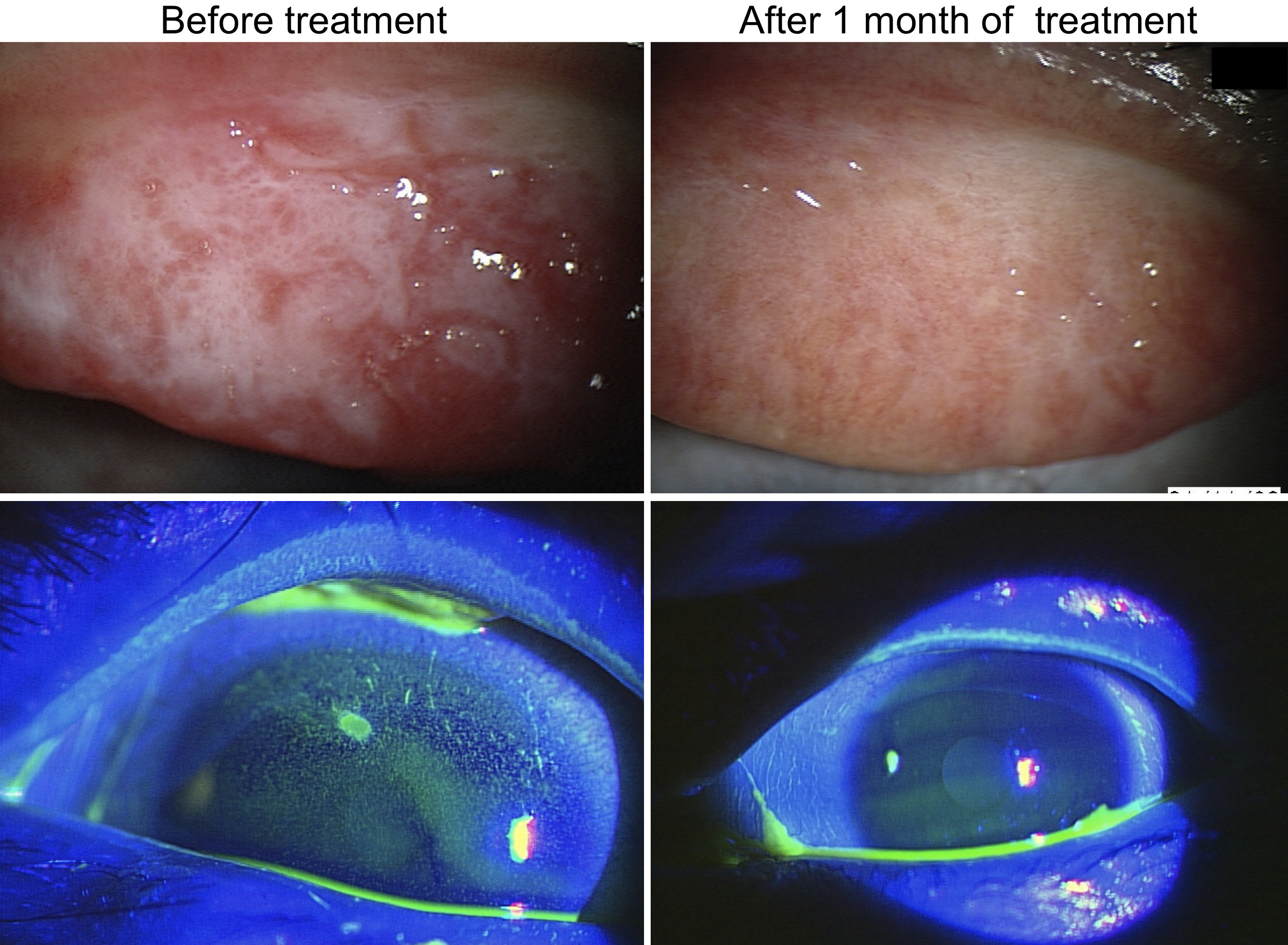
 Figure 1 of Wakamatsu, Mol Vis 2011; 17:932-938.