Figure 3. Dialyzing cones with 40 mM
glutamate increased the concentration of glutamate within the synaptic
cleft. A: Example of horizontal cell excitatory postsynaptic
currents (EPSCs) evoked by depolarizing stimulation of a presynaptic
cone without supplemental glutamate in control conditions (black
trace), in the presence of a low-affinity glutamate receptor antagonist
γDGG (0.1 mM, gray trace), and following washout (dashed trace). B:
Example
of horizontal cell EPSCs evoked by depolarizing stimulation of
a presynaptic cone dialyzed with 40 mM glutamate in control conditions
(black trace), during application of γDGG (gray trace), and following
washout (dashed trace). C: Bar graph showing the fractional
inhibition of EPSCs produced by 2.5 min. bath application of γDGG
(control, n=9; 40 mM, n=6; p=0.032, unpaired t-test) or the
high-affinity AMPA antagonist, NBQX (0.1 mM; control, n=8; 40 mM
glutamate, n=4; p=0.41, unpaired t-test). Error bars show SEM.
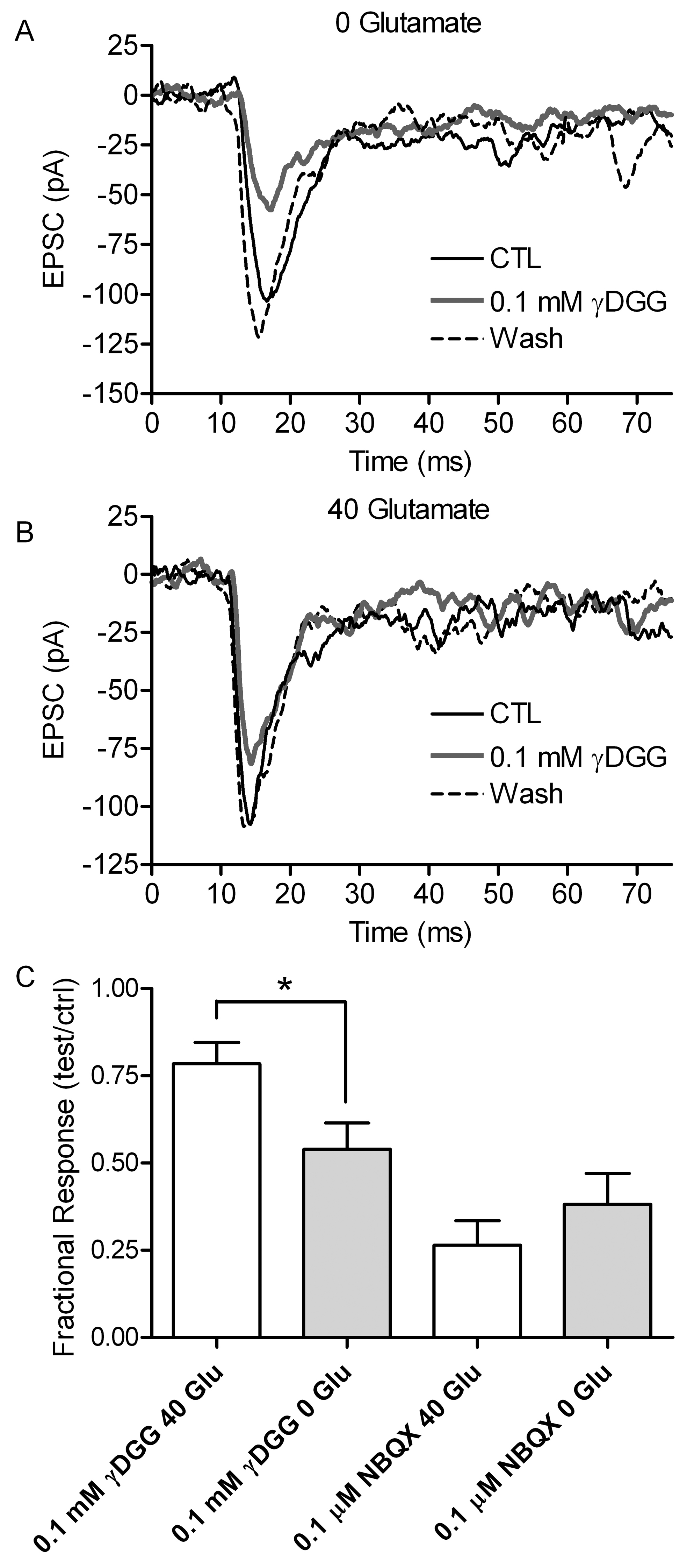
 Figure 3 of Bartoletti, Mol Vis 2011; 17:920-931.